. 2002 Mar 15;21(6):1289-300.
doi: 10.1093/emboj/21.6.1289.

Late endosome motility depends on lipids via the small GTPase Rab7

Affiliations

Late endosome motility depends on lipids via the small GTPase Rab7

Cécile Lebrand et al. EMBO J. .

Abstract

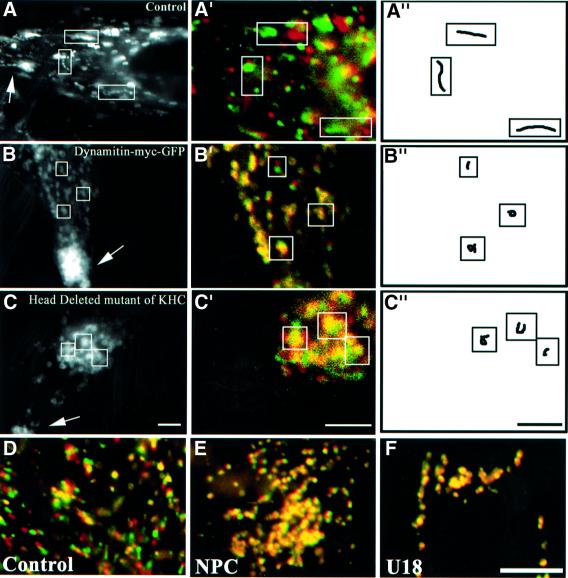
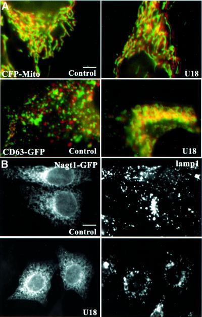
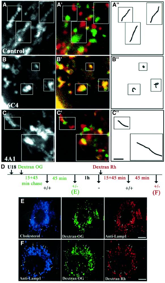
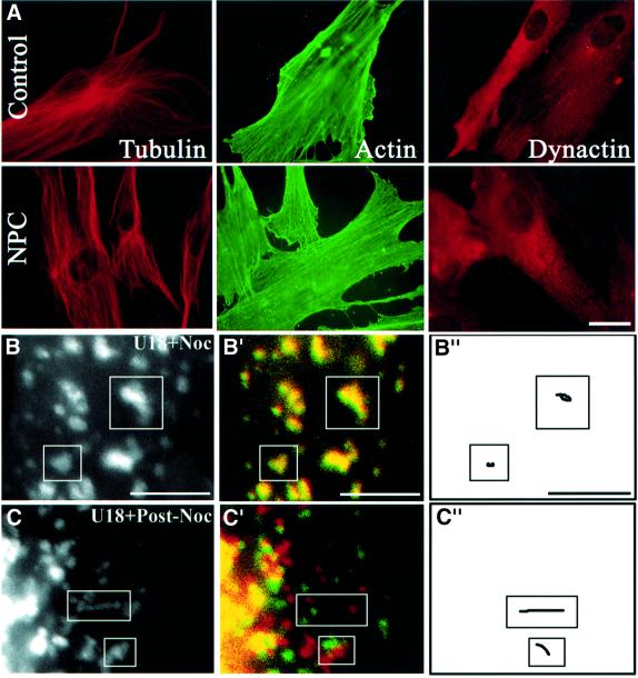
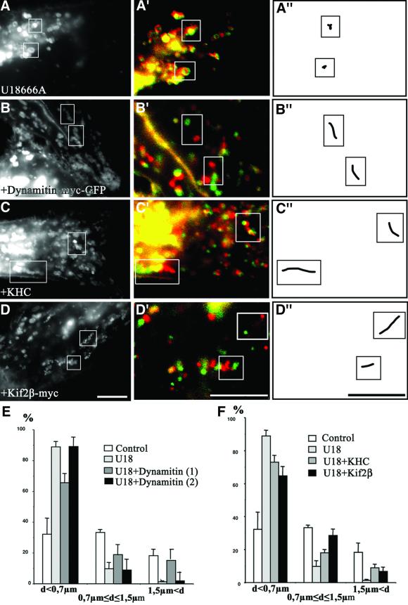
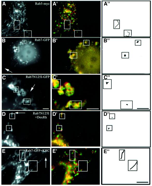
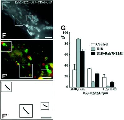
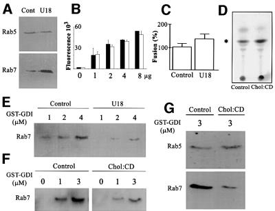
We report that lipids contribute to regulate the bidirectional motility of late endocytic compartments. Late endocytic vesicles loaded with cholesterol lose their dynamic properties, and become essentially immobile, including in cells from Niemann-Pick C patients. These vesicles then retain cytoplasmic dynein activity, but seem to be unable to acquire kinesin activity, eventually leading to paralysis. Our data suggest that this defect depends on the small GTPase Rab7, since the motility of vesicles loaded with cholesterol can be restored by the Rab7 inhibitory mutant N125I. Conversely, wild-type Rab7 overexpression mimics the effects of cholesterol on motility in control cells. Consistently, cholesterol accumulation increases the amounts of membrane-associated Rab7, and inhibits Rab7 membrane extraction by the guanine nucleotide dissociation inhibitor. Our observations thus indicate that cholesterol contributes to regulate the Rab7 cycle, and that Rab7 in turn controls the net movement of late endocytic elements. We conclude that motor functions can be regulated by the membrane lipid composition via the Rab7 cycle.


Figures

None
Fig. 1. Motility and cholesterol accumulation. (A–C) HeLa cells were transfected with CD63–GFP (A) or co-transfected with CD63–GFP and either dynamitin-myc–GFP (B), or a head-deleted mutant of KHC (C). Dynamitin-myc–GFP remained exclusively cytosolic, and was not detected after 100–200 ms exposure times; co-expression was verified by indirect immunofluorescence using anti-myc antibodies for dynamitin, or the Suk4 anti-kinesin antibody for the KHC mutant (endogenous and overexpressed KHC were easily distinguished by the intensity of the signal). Images were collected at 1 s interval over a time period of 25 s and then all images were stacked; arrows point to the cell periphery. Then, a moving object appears as a series of closely associated spots that reveals its track, as in the control cell in (A). (A′–C′) Initial and final positions were color-coded in red and green, respectively: a moving object is both red and green, and an immobile object yellow. (A′′–C′′) Traces of individual elements. Control (D and F) or NPC (E) fibroblasts were incubated at 37°C for 13 h with Alexa568-labeled antibodies against CD63 without (D and E) or with (F) 3 µg/ml U18666A, and analyzed as above (A′–C′). Bars: (A–C) 2 µm; (D–F) 3.7 µm.
None
Fig. 2. Other organelles. (A) HeLa cells transfected with the mitochondrial marker ECFP (CFP-mito) or with CD63–GFP were treated or not with U18666A. Frames were captured every 12 s to limit light damage, over 5 min to visualize better mitochondrial motility, and analyzed as in Figure 1A′–C′. (B) HeLa cells expressing NAGT1–GFP treated or not with U18666A and then with brefeldin A for 30 min, as indicated, were labeled with anti-Lamp1 antibodies and processed for microscopy. Bars: (A) 3.9 µm; (B) 6.2 µm.
None
Fig. 3. Antibodies against LBPA and pulse–chase. (A and B) HeLa or (C) BHK cells transfected with CD63–GFP were incubated for 24 h without (A) or with the monoclonal antibody against LBPA (B; 6C4) or Lamp1 (C; 4A1). (We used BHK cells because large amounts of 4A1 were available; cholesterol also accumulates in BHK cells after 6C4 treatment; Kobayashi et al., 1999.) Motility was analyzed as in Figure 1. (D) Outline. HeLa cells treated for 10 h with U18666A (the drug remained present throughout the experiment), were labeled with a 15 min pulse of endocytosed Oregon green–dextran (Dextran OG). After a 45 min chase, labeled endosomes were clearly motile (+/+). Motility was low after a second 45 min chase (+/–) and abolished after a third 1 h chase (–). Then, cells were labeled with a second pulse of rhodamine–dextran (Dextran Rh), and the chase protocol was repeated. Samples were fixed and analyzed by triple-channel fluorescence microscopy after the first (E) and second (F) wave. Bars: (A–C) 1.5 µm; (E) 4 µm; (F) 2.4 µm.
None
Fig. 4. Microtubules. (A) Control or NPC fibroblasts were labeled with the indicated antibodies. (B and C) HeLa cells transfected with CD63–GFP were treated for 13 h with U18666A, and the drug remained present throughout the experiment. Cells were then treated for 1 h with nocodazole (B; U18 + Noc) and then re-incubated for 90 min without nocodazole (C; U18 + Post-Noc). Motility analysis was as in Figure 1. Bars: (A) 8 µm; (B and C) 4 µm.
None
Fig. 5. Motors. Cells were transfected with CD63–GFP alone (A), or co-transfected with CD63–GFP and either dynamitin-myc–GFP (B), KHC (C) or Kif2β-myc (D). (As in Figure 1, overexpressed dynamitin remained cytosolic; co-expression was verified in all cases.) Cells were then treated with U18666A for 13 h. Motility analysis was as in Figure 1. Bars: 4 µm. (E and F) The bird’s eye distances (not the trajectory) between initial and final positions were quantified after 25 s. Control, control without U18666A; U18, U18666A as in (A); U18 + Dynamitin, dynamitin and U18666A; motility was partially restored as in (B) (1), but stopped at the cell periphery (2); U18 + KHC, KHC and U18666A as in (C); U18 +Kif2β, Kif2β and U18666A as in (D).
None
Fig. 6. Rab7 and cholesterol. (A) Co-transfection with CD63–GFP and Rab5-myc (co-expression was verified by immunofluorescence using anti-myc antibodies). (B) Transfection with Rab7–GFP. (C) Co-transfection with CD63–GFP and Rab7N125I–GFP (Rab7N125I–GFP remained cytosolic and was not detected after 100–200 ms exposure; co-expression was verified after longer exposures). (D) After transfection with Rab7N125I–GFP, late endocytic vesicles were labeled with a 15 min pulse of rhodamine–dextran (DexRh) followed by a 45 min chase, as in Figure 3D, to reveal better the partial redistribution to the periphery. (E) Cells co-transfected with Rab7–GFP and KHC were treated with U18666A for 13 h. (F and G) Cells co-transfected with CD63–GFP and Rab7N125I–GFP were treated with U18666A for 13 h (F), and distances between initial and final positions were quantified (G) as in Figure 5. Motility analysis was as in Figure 1. Arrows point to the cell periphery. Bars: (A, D and E) 4 µm; (B and C) 2 µm; (F) 2.8 µm.
None
Fig. 7. Extraction by GDI. (A and B) Since fractionation properties can be altered by cholesterol accumulation (Lange et al., 1998), we prepared crude fractions containing both early and late endosomes from control (cont) or U18666A-treated (U18) BHK cells, so that samples and Rab protein content could be compared directly in the same experiment. Fractions were analyzed by western blotting with antibodies against Rab5 or Rab7 (A) or by ELISA using the anti-LBPA antibody (B; increasing amounts of fractions). (C) Homotypic fusion of late endosomes from control or U18666A-treated BHK cells. Fusion activity was normalized to the control. (D) Crude fractions from control cells were loaded (chol:CD) or not (control) with cholesterol in vitro using 1 mM cholesterol complexed to methyl-β-cyclodextrin. After flotation on gradients, membranes were collected, and lipids analyzed by TLC; the position of cholesterol is indicated. (E) Cells were treated (U18) or not (control) with U18666A. Crude fractions were incubated with 1, 2 or 4 µM GST–GDI (Cavalli et al., 2001). [To avoid any bias, control (∼30 µg) and cholesterol-loaded membranes (∼15 µg) were normalized to Rab7 itself.] Endosomes were removed by flotation in gradients. GST–GDI with captured Rab7 was recovered onto glutathione beads. Beads were analyzed by western blotting with anti-Rab7 antibodies. (F) Membranes loaded or not with cholesterol in vitro as in (D) were analyzed as in (E). (G) As (F), except that 3 µM GST–GDI was used, and analysis was with antibodies against Rab5 or Rab7.

Similar articles

Cited by

References

    1. Allan V.J. and Schroer,T.A. (1999) Membrane motors. Curr. Opin. Cell Biol., 11, 476–482. - PubMed
    1. Aniento F., Emans,N., Griffiths,G. and Gruenberg,J. (1993) Cytoplasmic dynein-dependent vesicular transport from early to late endosomes [published erratum appears in J. Cell Biol. (1994) 124, 397]. J. Cell Biol., 123, 1373–1387. - PMC - PubMed
    1. Berrueta L., Tirnauer,J.S., Schuyler,S.C., Pellman,D. and Bierer,B.E. (1999) The APC-associated protein EB1 associates with components of the dynactin complex and cytoplasmic dynein intermediate chain. Curr. Biol., 9, 425–428. - PubMed
    1. Bomsel M., Parton,R., Kuznetsov,S.A., Schroer,T.A. and Gruenberg,J. (1990) Microtubule and motor dependent fusion in vitro between apical and basolateral endocytic vesicles from MDCK cells. Cell, 62, 719–731. - PubMed
    1. Bucci C., Thomsen,P., Nicoziani,P., McCarthy,J. and van Deurs,B. (2000) Rab7: a key to lysosome biogenesis. Mol. Biol. Cell., 11, 467–480. - PMC - PubMed

Publication types

MeSH terms

Substances