. 2002 Mar 15;21(6):1369-78.
doi: 10.1093/emboj/21.6.1369.

Structure-based analysis of RNA polymerase function: the largest subunit's rudder contributes critically to elongation complex stability and is not involved in the maintenance of RNA-DNA hybrid length

Affiliations

Structure-based analysis of RNA polymerase function: the largest subunit's rudder contributes critically to elongation complex stability and is not involved in the maintenance of RNA-DNA hybrid length

Konstantin Kuznedelov et al. EMBO J. .

Abstract

Analysis of multisubunit RNA polymerase (RNAP) structures revealed several elements that may constitute the enzyme's functional sites. One such element, the 'rudder', is formed by an evolutionarily conserved segment of the largest subunit of RNAP and contacts the nascent RNA at the upstream edge of the RNA-DNA hybrid, where the DNA template strand separates from the RNA transcript and re-anneals with the non-template strand. Thus, the rudder could (i) maintain the correct length of the RNA-DNA hybrid; (ii) stabilize the nascent RNA in the complex; and (iii) promote or maintain localized DNA melting at the upstream edge of the bubble. We generated a recombinant RNAP mutant that lacked the rudder and studied its properties in vitro. Our results demonstrate that the rudder is not required for establishment of the upstream boundary of the transcription bubble during promoter complex formation, nor is it required for separation of the nascent RNA from the DNA template strand or transcription termination. Our results suggest that the rudder makes critical contributions to elongation complex stability through direct interactions with the nascent RNA.


Figures
None
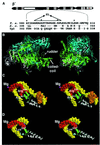
Fig. 1. Genetic and structural context of the RNAP rudder. (A) The bar at the top represents the largest RNAP subunit. The lettered boxes indicate evolutionarily conserved segments (Allison et al., 1985), the white box represents a long insertion in T.aquaticus β′ that is absent from the E.coli β′. The T.aquaticus segment C sequence (T. a.) is expanded underneath and is aligned with the corresponding segments from E.coli (E. c.), and yeast RNAP II (Yp2). The deletion studied in this work and the two amino acid linker (EL) inserted instead of the deleted material are shown above the T.aquaticus sequence. (B) The structure of T.aquaticus RNAP core enzyme (Zhang et al., 1999). The view on the left panel shows the ‘downstream face’ of the enzyme and is roughly parallel to the axis of the DNA-binding channel of the enzyme. Right panel: the view on the left was rotated 90° clockwise about the vertical axis. β′ is in green, β in cyan, α2 in white, ω in gray. The active center Mg2+ is in magenta. The β′ rudder and the coiled-coil element from which the rudder emanates are indicated. (C) The position of the β′ rudder according to the structural model of the bacterial RNAP elongation complex (Korzheva et al., 2000). A stereo view of the rudder area (roughly corresponding to the orientation shown in B, right panel) is shown. RNA is in red, template DNA strand in yellow, non-template DNA in orange (only the upstream portion of non-template DNA is shown, for clarity) and the active-center Mg2+ in magenta. The coiled-coil element is shown in gray and the rudder in green. Two evolutionarily conserved arginine residues of the rudder are indicated and are shown in spacefill representation. Conserved arginine residues located C-terminal to the rudder (A) are indicated in cyan. (D) The position of the rudder in the structure of the artificial elongation complex formed by yeast RNAP II (Gnatt et al., 2001). A stereo view of the relevant section of the yeast RNAP II elongation complex is shown. The orientation and labeling correspond to those in (C). A portion of the rudder is not resolved on this structure.
None
Fig. 2. Thermus aquaticus RNAP mutants harboring lesions in the β′ rudder form inactive promoter complexes. (A) Abortive transcription by the wild-type and mutant T.aquaticus RNAPs. The indicated enzymes were combined with the T7 A1 promoter-containing DNA fragment in the presence of CpA primer and [α-32P]UTP substrate. The reaction proceeded for 15 min at 65°C, and reaction products were resolved by denaturing PAGE and revealed by autoradiography. (B) Promoter complexes formed by the T.aquaticus rudder mutant are partially melted and are similar to complexes formed by E.coli RNAP with lesions in β dispensable region 1. A 170 bp DNA fragment containing the T7 A2 promoter (–83 to +87) 32P end-labeled on the bottom strand was combined with the indicated enzymes. Reactions were pre-incubated for 15 min at 37°C (E.coli RNAP), or for 15 min at 65°C (T.aquaticus RNAP), and probed with KMnO4. Reaction products were resolved on a 6% sequencing gel and visualized by autoradiography. On the right, phosphoimager traces of lanes 1, 2, 3 and 4 are presented. (C) Deletion of the rudder does not stabilize promoter complexes at suboptimal temperatures. The T7 A2 promoter complexes were formed at temperatures ranging from 5 to 75°C with 10°C increments and probed with KMnO4. Reaction products were analyzed as above.
None
Fig. 3. Transcription by T.aquaticus RNAP mutants from mismatched bubble templates. The indicated RNAP holoenzymes were combined with the T7 A1 promoter (–46 to +20) synthetic DNA fragment (lanes 1 and 5) or the T7 A1 promoter with the synthetic mismatched bubble templates indicated at the top of the figure. Reactions were supplemented with CpApUpC primer, 50 µM NTPs and [α-32P]CTP. The reaction proceeded for 10 min at 55°C, and reaction products were resolved by denaturing PAGE and revealed by autoradiography.
None
Fig. 4. Affinity labeling of T.aquaticus RNAP mutants on minimal scaffold templates. (A) Affinity labeling of RNAP β′ using a lysine-specific aldehyde-based reagent on the 5′ end of the nascent RNA. Artificial elongation complexes were assembled using the indicated RNAP core enzymes and the minimal nucleic acid scaffold templates schematically shown at the top of the figure. The 8mer RNA was elongated with radioactive CTP specified by the template. The position of the radioactive residue is indicated by an asterisk. The 5′ end of RNA was derivatized with a lysine-specific cross-linkable aldehyde group. Protein–RNA cross-links were induced by treatment with 10 mM NaBH4, and reaction products were resolved by SDS–PAGE and revealed by autoradiography. (B) The results of single-hit CNBr treatment of the major radioactive band from (A), lane 4, are shown. The distribution of N-terminal radioactively labeled CNBr fragments based on the known T.aquaticus β′ sequence is illustrated schematically on the right. M is a marker lane prepared using T.aquaticus β′ cross-linked to radioactively labeled RNA at an N-terminal site (labeled by an open circle on the schematics on the right).
None
Fig. 5. Determining the length of the RNA–DNA hybrid in the RNAP rudder mutant. (A) The experiment was performed as described in the text (see also Korzheva et al., 1998). Lanes 1, 5 and 9, RNA in initial complexes; lanes 2, 6 and 10, RNA in washed complexes that underwent limited pyrophosphorolysis. Lanes labeled β, β and DNA show RNAs cross-linked to the largest RNAP subunits and template DNA, respectively. (B) Quantification of the results in (A). The calculated efficiency of RNA–DNA cross-linking as a function of RNA length is presented. The height of the bars reflects the fraction of the RNA product cross-linked to DNA related to the total amount of the corresponding RNA in the complex (100% – RNA cross-linked to protein and DNA). (C) Elongation complexes stalled at position +11 were prepared using the indicated core RNAP enzymes and mismatched bubble templates fused to the λ tR2 terminator. Transcription was resumed by the addition of 250 µM NTPs. Reactions proceeded for 10 min at 55°C and were terminated by the addition of formamide-containing buffer (lanes 1 and 3). In lanes 2 and 4, 2.5 U of RNase H was added to completed transcription reactions for 10 min at 37°C prior to the addition of loading buffer. Lanes 5 and 6 are controls establishing that RNase H efficiently recognized products of in vitro transcription reactions (lane 5) annealed to a complementary DNA fragment, resulting in shorter RNA products (indicated by arrows in lane 6). The reaction products were resolved by denaturing PAGE and visualized by autoradiography.
None
Fig. 6. Transcription complexes formed by RNAP without the β′ rudder are unstable at elevated salt concentrations. (A) Transcription elongation in the presence of increasing concentrations of KCl. Stalled elongation complexes were formed as described in the legend of Figure 5C, and transcription was resumed by the addition of 250 µM NTPs and in the presence of the indicated concentration of KCl in the buffer. The reaction products were resolved by denaturing PAGE, visualized by autoradiography and quantified by phosphoimagery. (B) The rudder contributes to the ability of the transcription complex to withstand high concentrations of salt in the absence of the non-template DNA strand. Immobilized transcription complexes containing radioactively labeled nine nucleotide long RNA were assembled using scaffold template shown schematically at the top of the figure. The complexes were then transferred into a buffer containing 0.5 NaCl, incubated for the times indicated, and the amount of RNA remaining in the complex was revealed by denaturing PAGE and autoradiography. The gels were also quantified by phosphoimagery and the results are presented at the right of the figure. At the bottom of the figure, the results of a similar experiment conducted at low (40 mM) NaCl concentration in the buffer are presented.

Similar articles

Cited by

References

    1. Allison L.A., Moyle,M., Shales,M. and Ingles,C.J. (1985) Extensive homology among the largest subunits of eukaryotic and prokaryotic RNA polymerases. Cell, 42, 599–610. - PubMed
    1. Borukhov S. and Goldfarb,A. (1993) Recombinant Escherichia coli RNA polymerase: purification of individually overexpressed subunits and in vitro assembly. Protein Expr. Purif., 4, 503–511. - PubMed
    1. Burgess R.R., Arthur,T.M. and Pietz,B.C. (1998) Interaction of Escherichia coli σ70 with core RNA polymerase. Cold Spring Harb. Symp. Quant. Biol., 63, 277–287. - PubMed
    1. Chamberlin M.J. (1992) New models for the mechanism of transcription elongation and its regulation. Harvey Lect., 88, 1–21. - PubMed
    1. Cramer P., Bushnell,D.A. and Kornberg,R.D. (2001) Structural basis of transcription: RNA polymerase II at 2.8 Å resolution. Science, 292, 1863–1876. - PubMed

Publication types

MeSH terms