. 2002 Mar 15;21(6):1398-405.
doi: 10.1093/emboj/21.6.1398.

The Wilms' tumor gene Wt1 is required for normal development of the retina

Affiliations

The Wilms' tumor gene Wt1 is required for normal development of the retina

Kay-Dietrich Wagner et al. EMBO J. .

Abstract

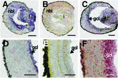
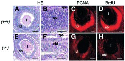
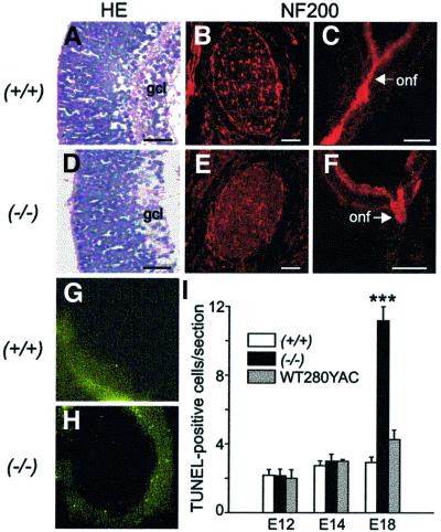
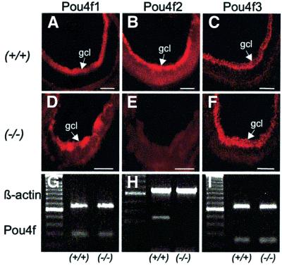
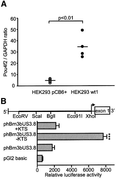
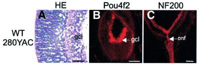
The Wilms' tumor gene Wt1 is known for its important functions during genitourinary and mesothelial formation. Here we show that Wt1 is necessary for neuronal development in the vertebrate retina. Mouse embryos with targeted disruption of Wt1 exhibit remarkably thinner retinas than age-matched wild-type animals. A large fraction of retinal ganglion cells is lost by apoptosis, and the growth of optic nerve fibers is severely disturbed. Strikingly, expression of the class IV POU-domain transcription factor Pou4f2 (formerly Brn-3b), which is critical for the survival of most retinal ganglion cells, is lost in Wt1(-/-) retinas. Forced expression of Wt1 in cultured cells causes an up-regulation of Pou4f2 mRNA. Moreover, the Wt1(-KTS) splice variant can activate a reporter construct carrying 5'-regulatory sequences of the human POU4F2. The lack of Pou4f2 and the ocular defects in Wt1(-/-) embryos are rescued by transgenic expression of a 280 kb yeast artificial chromosome carrying the human WT1 gene. Taken together, our findings demonstrate a continuous requirement for Wt1 in normal retina formation with a critical role in Pou4f2-dependent ganglion cell differentiation.


Figures

None
Fig. 1. Representative micrographs of tissue sections of wild-type (Wt1+/+) eyes at different stages of development. Wt1 transcripts are detected by mRNA in situ hybridization in the lens vesicle and retinal neuroblasts of E12 mice (A), as well as in the developing ganglion cell layer of E15 (C) and P1 eyes (D). No Wt1 mRNA is visible in the retinas of adult mice (E) and in retinal tissue of E12 embryos hybridized with digoxigenin-labeled Wt1 sense RNA strand (B). Note, that WT1 protein is detected by immunohistochemistry in the inner portion of the retina obtained at autopsy of a 19-week-gestation human embryo (F). l, lens; nbl, neuroblast layer; gcl, ganglion cell layer. Scale bars, 100 µm.
None
Fig. 2. Representative (immuno)histology of wild-type (+/+) and Wt1 null mutant (–/–) eyes at E12. HE staining reveals notably thinner retinas in Wt1–/– (E and F) than in wild-type embryos (A and B). Immunofluorescent labeling of PCNA is clearly reduced in the mutant (G) versus Wt1+/+ (C) retina. Incorporation of BrdU as an estimate of DNA synthesis is also decreased in Wt1–/– (H) compared with wild-type (D) eyes. l, lens; nbl, neuroblast layer; rpe, retinal pigment epithelium. Scale bars, 100 µm (A, C–E, G and H) and 50 µm (B and F), respectively.
None
Fig. 3. Morphology of wild-type (AC) and Wt1–/– mutant (DF) retinas at E18. A significant (∼40%) reduction in the number of ganglion cells is detected by HE staining in the E18 retinas of Wt1–/– (D) compared with wild-type (A) embryos. Immunofluorescent labeling of NF200 reveals blind-ending optic nerve fibers exiting from the Wt1–/– eyes (F). NF200 immunoreactivity is absent from cross-sections of optic nerves made at a distance of ∼400 µm beyond the optic disc level (E). For comparison, NF200-positive axon bundles are clearly visible in the optic nerves of wild-type retina (B). Shown are representative results from five different embryos in both groups. gcl, ganglion cell layer; onf, optic nerve fibers. Scale bars, 50 µm (A and D), 100 µm (B and E) and 200 µm (C and F). Representative examples of apoptotic cells identified by TUNEL assay in the E18 retinas of wild-type (G) and Wt1–/– (H) mice. The majority (∼60%) of the TUNEL-positive cells are located in the developing ganglion cell layer of the mutant retina (H). (I) The number of apoptotic cells identified by TUNEL assay in the neural retinas of wild-type (Wt1+/+), null mutant (Wt1–/–) and WT280-YAC transgenic embryos at different stages of development. An ∼4-fold increase in apoptotic cells is counted in mutant versus Wt1+/+ retinas at E18. Note that the average number of apoptotic cells in the developing retina is not significantly different between wild-type and WT280–YAC embryos. Five animals were studied in each group at the indicated time points. Five 10 µm retinal sections were made from each embryo close to the optic disc level. Values shown are means ± SEM. Asterisks indicate statistical significance (P <0.001, ANOVA with Bonferroni as post-hoc test).
None
Fig. 4. Expression of Pou4f genes in retinal ganglion cells of wild-type (+/+) and Wt1 null mutant (–/–) retinas at E18. Negative immuno fluorescent staining of the Wt1–/– retina (E) indicates lack of expression of the class IV POU-domain transcription factor Pou4f2. RT–PCR demonstrates absence of Pou4f2 mRNA from the E18 retinas of Wt1–/– embryos (H). No differences are seen in Pou4f1 and Pou4f3 expression (mRNA and protein) between wild-type and Wt1–/– retinas. Shown are representative data obtained from five different embryos each. gcl, ganglion cell layer. Scale bars, 100 µm. A 100 bp DNA ladder was used to estimate the sizes of the expected PCR products. The predicted lengths of the amplified targets are 274 bp (Pou4f1), 300 bp (Pou4f2), 229 bp (Pou4f3) and 642 bp (β-actin), respectively.
None
Fig. 5. (A) Expression of Pou4f2 in HEK293 cells stably transfected either with a Wt1(–KTS) expression construct or with the empty pCB6+ vector. Pou4f2 mRNA levels were quantified by real-time RT–PCR using the light cycler system (Roche Molecular Biochemicals) and normalized for GAPDH transcripts. Shown are the data obtained from four independent clones each of pCB6+ and Wt1(–KTS) transfected cells. Note that stable expression of Wt1(–KTS) increased Pou4f2 mRNA levels in HEK293 cells ∼8-fold. The horizontal bars indicate the mean values in each group. (B) Relative luciferase activities measured in the lysates of U2OS human osteosarcoma cells. U2OS cells were transiently co-transfected with phBrn3bUS3.8, Wt1 expression constructs encoding two different splice variants (+KTS/–KTS isoforms), and a cytomegalovirus-promoter driven β-galactosidase expression vector that was used for normalization of transfection efficiencies. Plasmid phBrn3bUS3.8 contained an ∼3.8 kb EcoRV–XhoI genomic sequence from the 5′-regulatory region of the human Pou4f2 gene (schematic drawing) in the pGL2 basic reporter vector. Values shown are means ± SEM of n = 7 experiments each performed in duplicate. P <0.05 was considered statistically significant (ANOVA).
None
Fig. 6. Rescue of retinal phenotype by YAC complementation of the human WT1 gene in Wt1–/– E18 embryos. Note that normal histology of the developing retina (A), as well as Pou4f2 immunoreactivity in the ganglion cell layer (B) and optic nerve fiber growth (C), are re-established by transgenic expression of a 280 kb YAC construct carrying the human WT1 gene in Wt1–/– embryos. The eyes of WT280– YAC transgenic embryos are indistinguishable by morphological means from their wild-type counterparts at stage E18. gcl, ganglion cell layer; onf, optic nerve fibers. Scale bars, 50 µm (A), 100 µm (B) and 200 µm (C), respectively.

References

    1. Armstrong J.F., Pritchard-Jones,K., Bickmore,W.A., Hastie,N.D. and Bard,J.B. (1993) The expression of the Wilms’ tumor gene, WT1, in the developing mammalian embryo. Mech. Dev., 40, 85–97. - PubMed
    1. Bennington J. and Beckwith,J. (1975) Tumors in the kidney, renal pelvis and ureter. In Atlas of Tumor Pathology. Series 2, fascile 12. Armed Forces Institute of Pathology, Washington, DC.
    1. Bickmore W.A. and Hastie,N.D. (1989) Aniridia, Wilms’ tumor and human chromosome 11. Ophthalmic Paediatr. Genet., 10, 229–248. - PubMed
    1. Braissant O. and Wahli,W. (1998) A simplified in situ hybridization protocol using non-radioactively labeled probes to detect abundant and rare mRNAs on tissue sections. Biochemica, 1, 10–16.
    1. Buckler A.J., Pelletier,J., Haber,D.A., Glaser,T. and Housman,D.E. (1991) Isolation, characterization and expression of the murine Wilms’ tumor gene (WT1) during kidney development. Mol. Cell. Biol., 11, 1707–1712. - PMC - PubMed

Publication types

MeSH terms