

Apoptosis and proliferation of acinar and islet cells in chronic pancreatitis: evidence for differential cell loss mediating preservation of islet function
- PMID: 11889077
- PMCID: PMC1773161
- DOI: 10.1136/gut.50.4.542
Apoptosis and proliferation of acinar and islet cells in chronic pancreatitis: evidence for differential cell loss mediating preservation of islet function
Abstract
Background: Chronic pancreatitis is characterised clinically by early exocrine insufficiency, with diabetes mellitus occurring as a late phenomenon. This is mirrored pathologically by extensive acinar cell destruction and islet preservation. The mechanisms underlying this differential rate of cellular destruction are unknown.
Aims: To test the hypothesis that acinar loss and islet preservation in chronic pancreatitis occurs due to differential epithelial kinetics and investigate the role of inflammatory cells and cell cycle associated molecules.
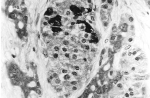
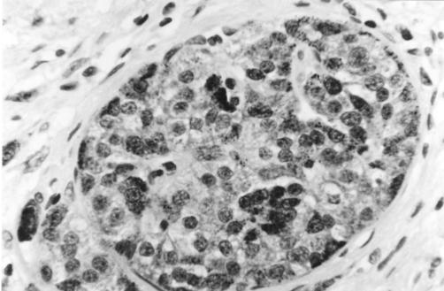
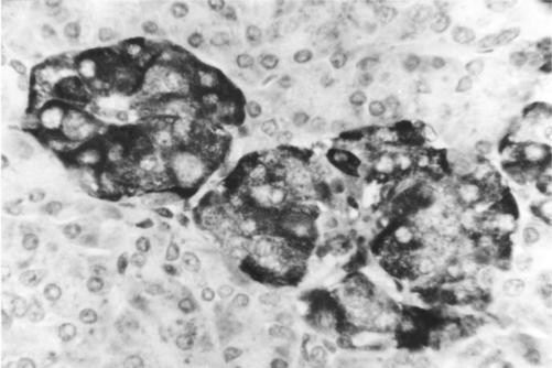
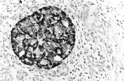
Methods: Archival tissue from six chronic pancreatitis cases was compared with six normal controls using TUNEL and immunohistochemistry for CD3, CD20, CD68, MIB-1, Bcl-2, Bax, Fas, Fas ligand, retinoblastoma protein (Rb), and tissue inhibitor of metalloproteinases 1 (TIMP-1) and 2 (TIMP-2).
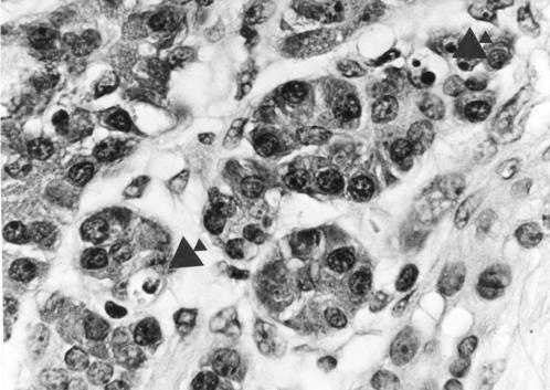
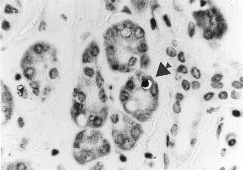
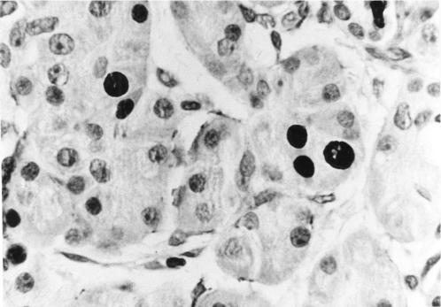
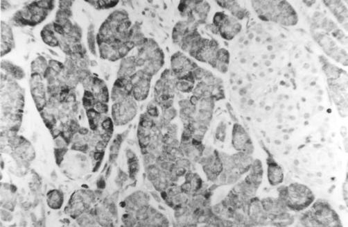
Results: The acinar cell apoptotic index (AI) and proliferation index were higher in chronic pancreatitis than controls. T lymphocytes diffusely infiltrated fibrous bands and acini but rarely islets. Acinar Bcl-2 expression exceeded islet expression in chronic pancreatitis and controls while Bax was strongly expressed by a subset of islet cells and weakly by centroacinar cells. Islet Fas and Fas ligand expression exceeded acinar expression in chronic pancreatitis and controls. Acinar Rb expression was higher in chronic pancreatitis than in controls. Islets in chronic pancreatitis and controls showed intense TIMP-1 and TIMP-2 expression.
Conclusion: Apoptosis plays a significant role in acinar loss in chronic pancreatitis. Acinar Bcl-2 and islet Bax expression indicates complex AI control. Increased acinar Rb expression in chronic pancreatitis may differentially promote acinar loss. Fas ligand expression may be restricted to islet cell membranes through TIMP-1 expression and inhibit islet damage by promoting apoptosis of cytotoxic T lymphocytes.
Figures
References
-
- Di Magno EP, Holtmann G. Chronic pancreatitis. Curr Opin Gastroenterol 1992;8:824–9.
-
- Kloppel G, Maillet B. Chronic pancreatitis: evolution of the disease. Hepatogastroenterology 1991;8:408–12. - PubMed
-
- Matthias PA, Ebert MD, Ademmer MD, et al. CD8+ CD103+ T-cells analogous to intestinal intraepithelial lymphocytes infiltrate the pancreas in chronic pancreatitis. Am J Gastroenterol 1998;93:2141–7. - PubMed
MeSH terms
Substances
LinkOut - more resources
Full Text Sources
Medical
Research Materials
Miscellaneous