. 2002 Apr;50(4):558-62.
doi: 10.1136/gut.50.4.558.

Attenuated acute liver injury in mice by naked hepatocyte growth factor gene transfer into skeletal muscle with electroporation

Affiliations

Attenuated acute liver injury in mice by naked hepatocyte growth factor gene transfer into skeletal muscle with electroporation

F Xue et al. Gut. 2002 Apr.

Abstract

Background: Hepatocyte growth factor (HGF) plays an essential role in hepatic development and regeneration, and shows proliferative and antiapoptotic activity in hepatocytes.

Aims: To establish an effective new method for HGF gene transfer in vivo and to investigate its effects in acute experimental liver injury.

Animals: Eight week old female mice were used.

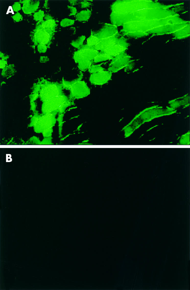
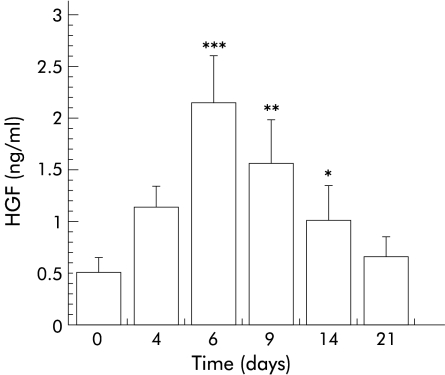
Methods: Rat HGF gene in a modified pKSCX plasmid was transferred to the tibialis anterior muscle by electroporation using a pulse generator. Four days later, plasma HGF concentrations were determined by enzyme linked immunosorbent assay every two days for three weeks. To confirm the efficacy of electroporation, a plasmid bearing green fluorescence protein (GFP) was transferred similarly. Four days after electroporation, carbon tetrachloride (CCl(4)) was administered to mice to induce acute liver injury. Plasma alanine aminotransferase (ALT) activity was measured. Hepatic apoptosis was assessed by Hoechst 33258 staining and the TUNEL method.

Results: Fluorescence microscopy showed strong green fluorescence where the GFP gene had been transferred into muscle. In mice given the HGF gene, HGF in plasma was increased up to fourfold from pretreatment amounts, peaking 6-9 days after electroporation and quickly decreasing within three weeks. Compared with the group without HGF transfer, the percentage of apoptotic hepatocytes after CCl(4) intoxication was significantly lower, as was ALT activity. In addition, ALT activity normalised more rapidly in the HGF gene transfer group.

Conclusions: Naked DNA injection and transfer by electroporation efficiently brings about HGF expression in vivo, which can attenuate acute liver injury.


Figures

Figure 1
Figure 1
Efficacy of gene transfer and expression by electroporation. Mice tibialis anterior muscle section was obtained seven days after electroporation with green fluorescence protein (GFP) plasmid. Homogeneous fluorescence was seen in the treated muscular fibres (A) but not in the control treated with phosphate buffered saline alone (B) (fluorescence microscopy, original magnification ×200).
Figure 2
Figure 2
Plasma levels of hepatocyte growth factor (HGF) at various time points. Blood samples were collected on days 4, 6, 9, 14, and 21 after electroporation with pKSCX-HGF plasmid and were detected by polyclonal antirat HGF ELISA. Data are expressed as mean (SD), n=10. *p<0.01, **p<0.005, ***p<0.001 versus control.
Figure 3
Figure 3
Nuclear morphology of hepatocytes two days after carbon tetrachloride intoxication. Deparaffinised section was stained with 8 μg/ml Hoechst 33258. Apoptotic nuclei were detected as condensed nuclei (A), DNA breaks (B, arrows), or marginally located at the nuclear membrane (B, arrowhead) (fluorescence microscopy, original magnification ×1000).
Figure 4
Figure 4
Apoptotic hepatocytes two days after carbon tetrachloride intoxication. Continuous liver sections were stained with haematoxylin and eosin (A) and TUNEL assay (B). (A) Apoptotic hepatocytes were shown adjacent to the central vein and were characterised by chromatin condensation (arrows) or apoptotic bodies (arrowheads). (B) The TUNEL positive nucleus was shown as condensed or DNA breaks (arrows). Necrotic cells exhibited a weakly non-specifically stained nucleus and cytoplasm (arrowhead) (microscopy, original magnification ×500).
Figure 5
Figure 5
TUNEL positive hepatocytes were evaluated on days 1, 2, 3, and 5 after administration of carbon tetrachloride (CCl4) in both hepatocyte growth factor (HGF) treated and control groups. TUNEL positive nuclei were counted in three random areas for each section, each with 4.4×105 pixels of area. Results are expressed as per cent apoptotic cells of the total number of hepatocytes in these fields. Data are means (SD). *p<0.01, **p<0.005 versus control.
Figure 6
Figure 6
Serial changes in proliferative cell nuclear antigen (PCNA) positive hepatocytes in hepatocyte growth factor (HGF) treated and control groups. PCNA positive nuclei were counted and expressed as a percentage of the total number of hepatocyte nuclei. A significant difference was observed only on day 1 after liver intoxication (*p<0.05).
Figure 7
Figure 7
Plasma alanine aminotransferase (ALT) activities at various times after intoxication with carbon tetrachloride (CCl4). Blood samples were detected by the dry chemical method using test paper. Data are expressed as mean (SD). *p<0.01, **p<0.005 versus the control group.

Similar articles

Cited by

References

    1. Nakamura T, Teramoto H, Ichihara A. Purification and characterization of a growth factor from rat platelets for mature parenchymal hepatocytes in primary cultures. Proc Natl Acad Sci USA 1986; 83:6489–93. - PMC - PubMed
    1. Nakamura T, Nawa K, Ichihara A, et al. Purification and subunit structure of hepatocyte growth factor from rat platelets. FEBS Lett 1987;224:311–16. - PubMed
    1. Matsumoto K, Nakamura T. Hepatocyte growth factor: Molecular structure, roles in liver regeneration, and other biological functions. Crit Rev Oncog 1992;3:27–54. - PubMed
    1. Boros P, Miller CM. Hepatocyte growth factor: a multifunctional cytokine. Lancet 1995;345:293–5. - PubMed
    1. Ricci G, Catizone A, Innocenzi A, et al. Hepatocyte growth factor (HGF) receptor expression and role of HGF during embryonic mouse testis development. Dev Biol 1999;216:340–7. - PubMed

MeSH terms