

Review
doi: 10.1136/gut.50.4.571.
Hepatic stellate cells: role in microcirculation and pathophysiology of portal hypertension
Affiliations
- PMID: 11889082
- PMCID: PMC1773176
- DOI: 10.1136/gut.50.4.571
Item in Clipboard
Review
Hepatic stellate cells: role in microcirculation and pathophysiology of portal hypertension
Gut.
2002 Apr.
Abstract
Accumulating evidence suggests that stellate cells are involved in the regulation of the liver microcirculation and portal hypertension. Activated hepatic stellate cells have the necessary machinery to contract or relax in response to a number of vasoactive substances. Because stellate cells play a role in both fibrosis and portal hypertension, they are currently regarded as therapeutic targets to prevent and treat the complications of chronic liver disease.
Figures
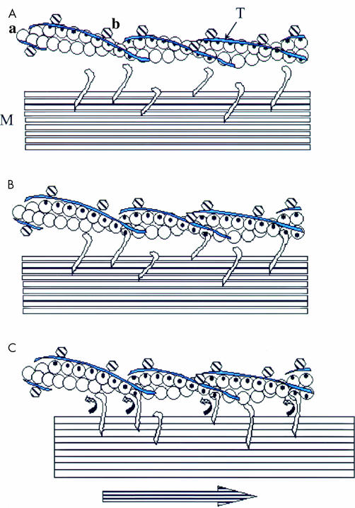
(A) Thin actin filament (a) and thick myosin filament (M) at rest, with tropomyosin (T) covering the myosin binding site and binding proteins with tropomyosin (b). In skeletal muscle, b is troponin; in smooth muscle it is likely to be calponin or caldesmon. (B) Activation of actomyosin: tropomyosin moves from myosin binding sites on actin in two stages—in skeletal muscle, stage 1 is caused by calcium binding to troponin while in smooth muscle stage 1 can be caused by myosin phosphorylation but that is not obligatory. In both cases initial myosin binding causes cooperative conformational changes in stage 2 to fully expose the actin binding sites for myosin. (C) Resulting actomyosin ATPase gives energy for a conformational change in myosin that moves the thick filament relative to the actin thin filament.
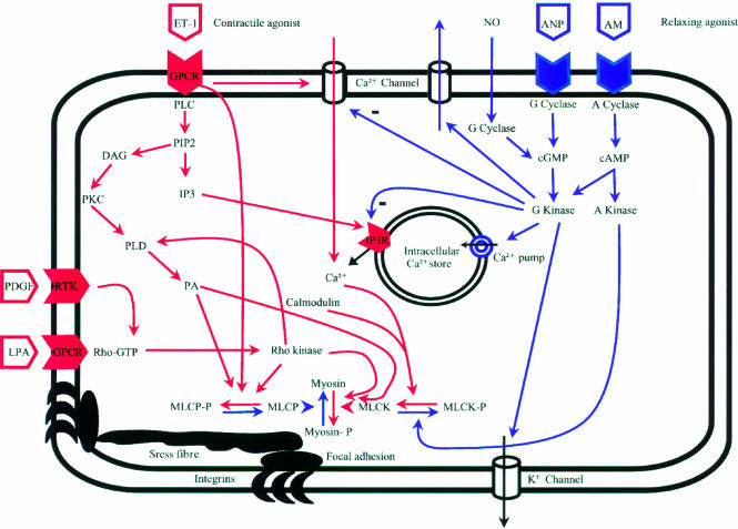
Schematic representation of intracellular contractile and relaxation mechanisms in hepatic stellate cells. It should be noted that most of the pathways have been described in smooth muscle cells but not all mechanism have been proved to exist in stellate cells. Pathways for contraction are marked in red whereas blue represents relaxation pathways. AM, adrenomedullin; ANP, atrial natriuretic peptide; DAG, diacylglycerol; ET-1, endothelin 1; GPCR, G protein coupled receptor; IP3, inositol 1,4,5-trisphosphate; LPA, lysophosphatidic acid; NO, nitric oxide; MLCK, myosin light chain kinase; MLCP, myosin light chain phosphatase; PIP2, phosphatidylinositol 4,5-bisphosphate; PLC, phospholipase C; PLD, phospholipase D; PKC, protein kinase C; RTK, receptor tyrosine kinase.
References
-
- Gupta TK, Chen L, Groszmann RJ. Pathophysiology of portal hypertension. Baillieres Clin Gastroenterol 1997;11:203–19. - PubMed
-
- Bhathal PS, Grossmann HJ. Reduction of the increased portal vascular resistance of the isolated perfused rat liver by vasodilators. J Hepatol 1985;1:325–37. - PubMed
-
- Ramadori G. The stellate cell of the liver. Virchows Arch B Cell Pathol 1991;61:147–58. - PubMed
Publication types
MeSH terms
Substances
LinkOut - more resources
Full Text Sources
Other Literature Sources