. 2002 Apr;184(7):1865-72.
doi: 10.1128/JB.184.7.1865-1872.2002.

Molecular analysis of the gene encoding a novel chitin-binding protease from Alteromonas sp. strain O-7 and its role in the chitinolytic system

Affiliations
Free PMC article

Molecular analysis of the gene encoding a novel chitin-binding protease from Alteromonas sp. strain O-7 and its role in the chitinolytic system

Katsushiro Miyamoto et al. J Bacteriol. 2002 Apr.
Free PMC article

Abstract

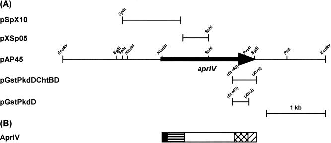
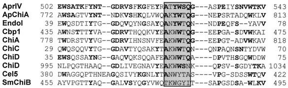
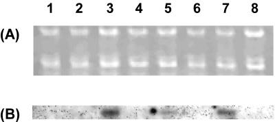
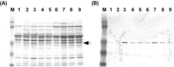
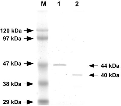
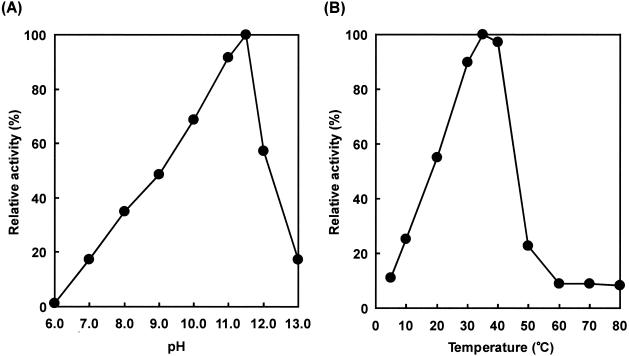
Alteromonas sp. strain O-7 secretes several proteins in response to chitin induction. We have found that one of these proteins, designated AprIV, is a novel chitin-binding protease involved in chitinolytic activity. The gene encoding AprIV (aprIV) was cloned in Escherichia coli. DNA sequencing analysis revealed that the open reading frame of aprIV encoded a protein of 547 amino acids with a calculated molecular mass of 57,104 Da. AprIV is a modular enzyme consisting of five domains: the signal sequence, the N-terminal proregion, the family A subtilase region, the polycystic kidney disease domain (PkdD), and the chitin-binding domain type 3 (ChtBD3). Expression plasmids coding for PkdD or both PkdD and ChtBD (PkdD-ChtBD) were constructed. The PkdD-ChtBD but not PkdD exhibited strong binding to alpha-chitin and beta-chitin. Western and Northern analyses demonstrated that aprIV was induced in the presence of N-acetylglucosamine, N-acetylchitobiose, or chitin. Native AprIV was purified to homogeneity from Alteromonas sp. strain O-7 and characterized. The molecular mass of mature AprIV was estimated to be 44 kDa by sodium dodecyl sulfate-polyacrylamide gel electrophoresis. The optimum pH and temperature of AprIV were pH 11.5 and 35 degrees C, respectively, and even at 10 degrees C the enzyme showed 25% of the maximum activity. Pretreatment of native chitin with AprIV significantly promoted chitinase activity.


Figures

FIG. 1.
FIG. 1.
Restriction map of pAP45 and domain structure of AprIV. (A) Restriction map of pAP45. Arrow indicates the ORF and the direction of transcription. aprIV, the gene encoding serine protease. (B) Diagram of domain structure of AprIV: signal peptide, ▪; N-terminal proregion, ▤; protease region, □; PkdD, ▩; ChtBD, ▨.
FIG. 2.
FIG. 2.
Comparison of the amino acid sequence of ChtBD of AprIV with those of other proteins. ApChiA, Aeromonas punctata chitinase A; EndoI, Vibrio furnissii chitodextrinase; Cbp1, ChiA, ChiC, and ChiD, chitin-binding protein, chitinase A, chitinase C, and chitinase-like enzyme from Alteromonas sp. strain O-7, respectively; Cel5, Erwinia chrysanthemi cellulase; SmChiB, Serratia marcescens chitinase B. The stWWst motif, where “s” and “t” represent small residues and turn-like residues, respectively, is shown by a shadowed box. Residues that are identical are indicated by bold letters.
FIG. 3.
FIG. 3.
Northern blot analysis of the aprIV transcript. Lanes: 1, control; 2 and 3, 0.5 and 1.0% GlcNAc, respectively; 4 and 5, 0.5 and 1.0% N-acetylchitobiose, respectively; 6, 0.5% glycol chitin; 7, 0.5% powdered chitin; 8, 1.0% skim milk. (A) Formaldehyde-agarose gel electrophoresis of the total RNA (1.2 μg) from the strain; (B) Northern blot analysis. The HindIII-SphI fragment (0.8 kb) from pAP45 was used as a probe.
FIG. 4.
FIG. 4.
Western blot analysis of AprIV. (A) Coomassie blue staining. Lanes: M, prestained molecular weight marker; 1, control; 2 and 3, 0.5 and 1.0% GlcNAc, respectively; 4 and 5, 0.5 and 1.0% N-acetylchitobiose, respectively; 6, 0.5% glycol chitin; 7, 8, and 9, 0.2, 0.5, and 1.0% powdered chitin, respectively. The arrow indicates AprIV protein. (B) Western blot analysis.
FIG. 5.
FIG. 5.
SDS-PAGE of AprIV and AprIVΔC. Lanes: M, marker proteins; 1, AprIV; 2, AprIVΔC.
FIG. 6.
FIG. 6.
Effects of pH and temperature on protease activity of AprIV. (A) The following buffer systems were used: 50 mM acetate buffer (pH 6.0), 50 mM Tris-HCl buffer (pH 7.0 to 9.0), and 50 mM glycine-NaOH buffer (pH 10.0 to 13.0). The reaction mixtures were incubated at 35°C for 30 min. The amount enzyme was 13.6 U. (B) The reaction was carried out at the various temperatures for 30 min at pH 11.5 (50 mM glycine-NaOH buffer). The amount of enzyme was 13.6 U.
FIG. 7.
FIG. 7.
Particitation of AprIV and AprIVΔC in chitin degradation. (A) Protease activities of AprIV and AprIVΔC were measured with the substrates native chitin (open bar) and deproteinized chitin (solid bar). Control experiments were carried out without enzyme. (B and C) The effects of AprIV and AprIVΔC on chitinase activity were examined by using native chitin (B) or deproteinized chitin (C). Symbols: •, Chi85; ▴, Chi85 plus AprIVΔC; ▪, Chi85 plus AprIV. Aliquots were taken at the indicated times and centrifuged, and chitinase activity was measured. The value after incubation for 6 h with AprIV and Chi85 was taken as 100%. Data from three independent experiments are shown; standard deviations are indicated by vertical lines.
FIG. 8.
FIG. 8.
Determination of the transcription start site of aprIV. (A) Primer extension and nucleotide sequencing were performed with the same FITC-labeled primer. The nucleotide sequence around the transcription start site is shown in lanes A, C, G, and T. The transcriptional start site is shown by an arrow (lane P). (B) Nucleotide sequence of the 5′ upstream regions of aprIV. The deduced amino acid sequence of AprIV is shown below the nucleotide sequence. The transcriptional start site is indicated by “+1.” The position of FITC-labeled primer is shown below the nucleotide sequence by an arrow. The putative −35 and −10 regions are shown by boxes. The conserved sequences are shown by shadowed boxes. The inverted repeat sequences are indicated by convergent arrows.

Similar articles

Cited by

References

    1. Ahsan, M. M., T. Kimura, S. Karita, K. Sakka, and K. Ohmiya. 1996. Cloning, DNA sequencing, and expression of the gene encoding Clostridium thermocellum cellulase CelJ, the largest catalytic component of the cellulosome. J. Bacteriol. 178:5732-5740. - PMC - PubMed
    1. Bradford, M. M. 1976. A rapid and sensitive method for the quantitation of microgram quantities of protein utilizing the principle of protein-dye binding. Anal. Biochem. 72:248-254. - PubMed
    1. Brun, E., F. Moriaud, P. Gans, M. J. Blackledge, F. Barras, and D. Marion. 1997. Solution structure of the cellulose-binding domain of the endoglucanase Z secreted by Erwinia chrysanthemi. Biochemistry 36:16074-16086. - PubMed
    1. Bycroft, M., A. Bateman, J. Clarke, S. J. Hamill, R. Sandford, R. L. Thomas, and C. Chothia. 1999. The structure of a PKD domain from polycystin-1: implications for polycystic kidney disease. EMBO J. 18:297-305. - PMC - PubMed
    1. Danielli, A., T. G. Loukeris, M. Lagueux, H. M. Muller, A. Richman, and F. C. Kafatos. 2000. A modular chitin-binding protease associated with hemocytes and hemolymph in the mosquito Anopheles gambiae. Proc. Natl. Acad. Sci. USA 97:7136-7141. - PMC - PubMed

LinkOut - more resources