

Antemortem MRI findings correlate with hippocampal neuropathology in typical aging and dementia
- PMID: 11889239
- PMCID: PMC2745935
- DOI: 10.1212/wnl.58.5.750
Antemortem MRI findings correlate with hippocampal neuropathology in typical aging and dementia
Abstract
Objectives: To assess the diagnostic specificity of MRI-defined hippocampal atrophy for AD among individuals with a variety of pathologically confirmed conditions associated with dementia as well as changes attributable to typical aging, and to measure correlations among premortem MRI measurements of hippocampal atrophy, mental status examination performance, and the pathologic stage of AD.
Methods: An unselected series of 67 individuals participating in the Mayo Alzheimer's Disease Research Center/Alzheimer's Disease Patient Registry who had undergone a standardized antemortem MRI study and also postmortem examination were identified. Hippocampal volumes were measured from antemortem MRI. Each postmortem specimen was assigned a pathologic diagnosis and in addition, the severity of AD pathology was staged using the method of Braak and Braak.
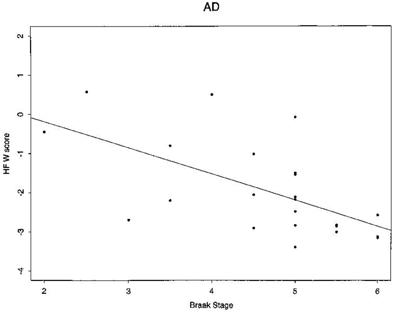
Results: Individuals with an isolated pathologic diagnosis of AD, hippocampal sclerosis, frontotemporal degeneration, and neurofibrillary tangle--only degeneration usually had substantial hippocampal atrophy, while those with changes of typical aging did not. Among all 67 subjects, correlations (all p < 0.001) were observed between hippocampal volume and Braak and Braak stage (r = -0.39), between hippocampal volume and Mini-Mental State Examination (MMSE) score (r = 0.60), and between MMSE score and Braak and Braak stage (r = -0.41).
Conclusions: Hippocampal atrophy, while not specific for AD, was a fairly sensitive marker of the pathologic AD stage (particularly among subjects with isolated AD pathology [r = -0.63, p = 0.001]) and consequent cognitive status.
Figures
References
-
- Braak H, Braak E. Neuropathological staging of Alzheimer-related changes. Acta Neuropathol. 1991;82:239–259. - PubMed
-
- Consensus recommendations for the postmortem diagnosis of Alzheimer's disease. The national Institute on Aging, and Reagan Institute Working Group on Diagnostic Criteria for the Neuropathological Assessment of Alzheimer's Disease. Neurobiol Aging. 1997;18:S1–S2. - PubMed
-
- de Leon MJ, Golomb J, Convit A, et al. Measurement of medial temporal lobe atrophy in diagnosis of Alzheimer's disease. The Lancet. 1993;341:125. - PubMed
-
- Fox NC, Warrington EK, Freeborough PA, et al. Presymptomatic hippocampal atrophy in Alzheimer's disease. A longitudinal MRI study. Brain. 1996;119:2001–2007. - PubMed
-
- Jack CR, Jr, Petersen RC, O'Brien PC, et al. MR-based hippocampal volumetry in the diagnosis of Alzheimer's disease. Neurology. 1992;42:183–188. - PubMed
Publication types
MeSH terms
Grants and funding
LinkOut - more resources
Full Text Sources
Medical