. 2002 Feb;13(2):95-101.
doi: 10.1007/s00335-001-2101-3.

Molecular characterization and mapping of ATOH7, a human atonal homolog with a predicted role in retinal ganglion cell development

Affiliations

Molecular characterization and mapping of ATOH7, a human atonal homolog with a predicted role in retinal ganglion cell development

Nadean L Brown et al. Mamm Genome. 2002 Feb.

Abstract

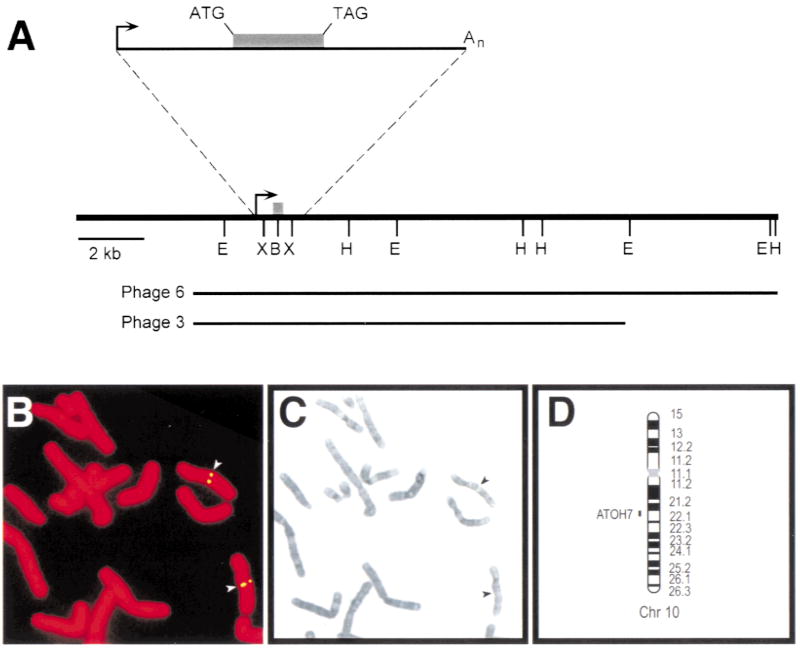
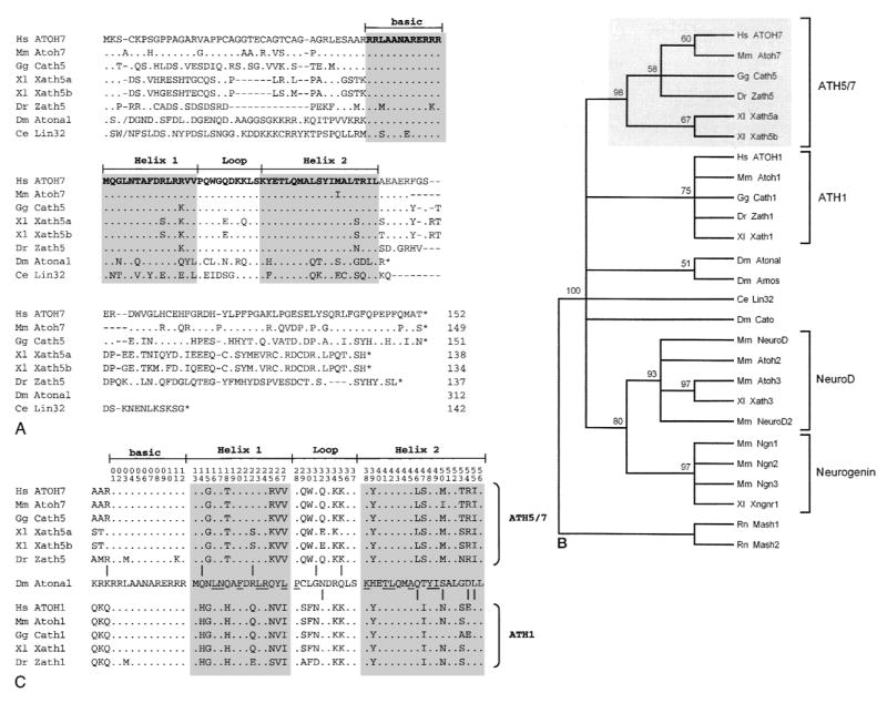
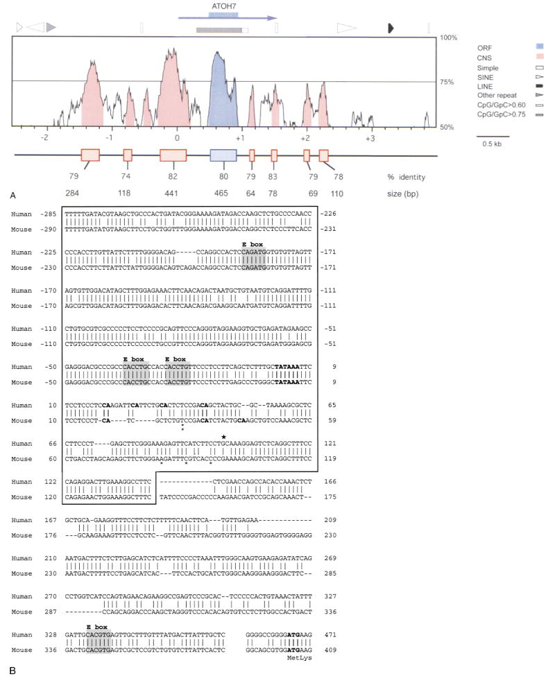
The human ATOH7 gene encodes a basic helix-loop-helix (bHLH) transcription factor that is highly similar to Drosophila Atonal within the conserved bHLH domain. The ATOH7 coding region is contained within a single exon. We mapped ATOH7 to Chromosome (Chr) 10q21.3-22.1, a region syntenic to the segment of mouse Chr 10 where Atoh7 (formerly Math5) is located. The evolutionary relationship between ATOH7 and other atonal homologs was investigated using parsimony analysis. A direct comparison of ATH5/7 and ATH1 protein subgroups to Atonal also revealed a nonrandom distribution of amino acid changes across the bHLH domain, which may be related to their separate visual and proprioceptive sensory functions. Among bHLH genes, ATOH7 is most closely related to Atoh7. This sequence conservation extends significantly beyond the coding region. We define blocks of strong homology in flanking human and mouse genomic DNA, which are likely to include cis regulatory elements. Because targeted deletion of Atoh7 causes optic nerve agenesis in mice, we propose ATOH7 as a candidate for human optic nerve aplasia and related clinical syndromes.


Figures

Fig. 1
Fig. 1
Map of ATOH7 and localization to human Chr 10q21.3–22.1 A. Restriction map derived from phage clones 3 and 6. The shaded box denotes the coding region, and the arrow indicates the start of transcription. Clone 6 was used for FISH analysis. The cDNA clone is represented by the solid line in the expanded view, and is coextensive with the genomic sequence. E, EcoRI; B, BamHI; H, HindIII; X, XhoI. B. Hybridization of biotin-labeled ATOH7 probe to propidium iodide-stained human female metaphase chromosomes. The signal was visualized indirectly with FITC. C. Giemsa-trypsin banding of the same metaphase spread. D. Ideogram showing the cytogenetic location of ATOH7 in 10q21.3–22.1.
Fig. 2
Fig. 2
Amino acid alignment and evolutionary tree showing ATOH7 within the Atonal bHLH family. A. Optimal alignment of full-length bHLH proteins. Amino acid identities to ATOH7, sequence gaps, and stop codons are indicated by periods, dashes, and asterisks, respectively. The slashes (/) near the N-termini of Atonal and Lin32 mark the positions where 196 and 31 amino acids were omitted, respectively. Lin32 was recently found to extend further upstream from the bHLH domain (Portman and Emmons 2000). B. Bootstrap majority rule consensus tree of the Atonal family. The tree was generated by comparing 26 bHLH domains (56 characters) using maximum parsimony analysis, with Mash1 and Mash2 as outgroup taxa. Above each branchpoint, we show the proportion of bootstrap replications (out of 100) that support the group descending from that node. A value over 70 indicates very strong support (Hillis and Bull 1993). The Atonal-Amos group is weakly supported. This phylogram is completely congruent with the strict consensus tree generated by a heuristic search. C. Distribution of 10 informative amino acid sites across the bHLH domain, where proteins in the ATH5/7 (top) and ATH1 (bottom) groups differ from each other, but where at least one member of the ATH5/7 or ATH1 groups is identical to Atonal. Amino acid positions are numbered at the top. The periods indicate identity with Atonal. Underlined residues are predicted to form intra- or intermolecular helical contacts (Atchley et al. 2000; Ferré-D’Amaré et al. 1993). These informative changes are nonrandomly distributed (Mann-Whitney rank test, US = 29, P = 0.005).
Fig. 3
Fig. 3
Extended DNA sequence similarity between human ATOH7 and mouse Atoh7. A. Distribution of human-mouse sequence homology for a DNA segment extending 3000 bp from each side of the ATOH7 open reading frame (ORF, blue box). The sequence is numbered in relation to the human transcription unit (blue arrow), starting with the conserved TATAAA box. The profile was created using VISTA and plots the percentage nucleotide identity (y-axis) calculated for a moving 100 bp window centered at each position of the aligned sequence (x-axis). Eight conserved nucleotide segments (CNS, shaded in red) were found in noncoding DNA within which all 100 bp windows are ≥70% identical. SINE, LINE, simple (low complexity) and other (MER) repetitive elements and CpG-rich regions in the human DNA are indicated above the graph. The boxes below the graph show highly conserved DNA segments revealed by pairwise BLAST2N analysis and the size and percentage identity of each alignment. Because of introduced gaps, the alignments are typically longer than the corresponding sequence in each species. In human DNA, the conserved segments extend from −1474 to −1193, −816 to −701, −285 to +142, +465 to +923 (coding), +1126 to +1189, +1438 to +1515, +1963 to +2031, and +2189 to +2297. B. Conservation of the promoter region. ATOH7 and Atoh7 sequences directly 5′ to the coding region are aligned. Vertical lines and dashes indicate nucleotide identity and gaps, respectively. The box encloses a 441-bp segment, revealed by BLASTN2, with 82% identity. The 5′ ends of the one human (★) and five mouse (*) cDNA clones (Brown et al. 1998) are shown above and below the aligned sequences, respectively. The TATAAA box, translational start site (ATG), and all potential transcription initiator (m7G cap) sites (CA) located 15–45 bp from the TATAAA box are indicated in bold type. The four conserved E box binding sites are shaded.

References

    1. Alagramam KN, Murcia CL, Kwon HY, Pawlowski KS, Wright CG, et al. The mouse Ames waltzer hearing-loss mutant is caused by mutation of Pcdh15, a novel protocadherin gene. Nat Genet. 2001;27:99–102. - PubMed
    1. Atchley WR, Wollenberg KR, Fitch WM, Terhalle W, Dress AW. Correlations among amino acid sites in bHLH protein domains: an information theoretic analysis. Mol Biol Evol. 2000;17:164–178. - PubMed
    1. Bermingham NA, Hassan BA, Price SD, Vollrath MA, Ben-Arie N, et al. Math1: an essential gene for the generation of inner ear hair cells. Science. 1999;284:1837–1841. - PubMed
    1. Bermingham NA, Hassan BA, Wang VY, Fernandez M, Banfi S, et al. Proprioceptor pathway development is dependent on math1. Neuron. 2001;30:411–422. - PubMed
    1. Bolz H, von Brederlow B, Ramirez A, Bryda EC, Kutsche K, et al. Mutation of CDH23, encoding a new member of the cadherin gene family, causes Usher syndrome type 1D. Nat Genet. 2001;27:108–112. - PubMed

Publication types

Associated data