

Review
doi: 10.1016/s0955-0674(02)00310-1.
Regulation of cell polarity during eukaryotic chemotaxis: the chemotactic compass
Affiliations
- PMID: 11891119
- PMCID: PMC2819118
- DOI: 10.1016/s0955-0674(02)00310-1
Item in Clipboard
Review
Regulation of cell polarity during eukaryotic chemotaxis: the chemotactic compass
Curr Opin Cell Biol.
2002 Apr.
Abstract
Phosphatidylinositol 3-kinase lipid products and the Rho GTPases play a central role in transmitting information from chemotactic receptors to the effectors of cell polarity, and recent advances in the field have allowed us to understand these roles more clearly. Emergent properties of positive and negative regulation of these molecules may account for the establishment of cell polarity during chemotaxis for a wide range of cells from Dictyostelium to fibroblasts to neutrophils.
Figures
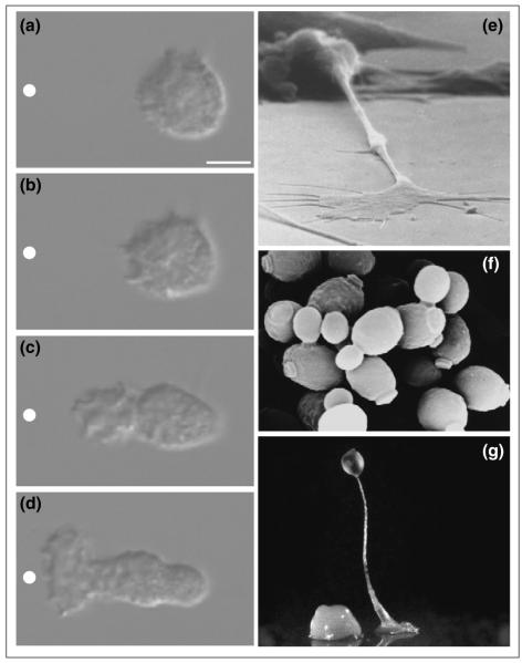
Examples of directed cell polarity. (a–d) Polarization of neutrophil in response to gradient of chemoattractant. Nomarski images of unpolarized neutrophil responding to a micropipette containing the chemoattractant FMLP (white circle) at (a) 5 s, (b) 30 s, (c) 81 s and (d) 129 s. Bar = 5μm. Figure reprinted from [3], with permission. (e) Similar processes are required for axons to find their way in the developing nervous system (courtesy of Ken Balazovich and Kathryn Tosney), (f) for Saccharomyces cerevisiae to bud and mate (courtesy of Angela Dunn and Mick Tuite), and (g) for Dictyostelium to form multicellular aggregates (courtesy of Rob Kay).
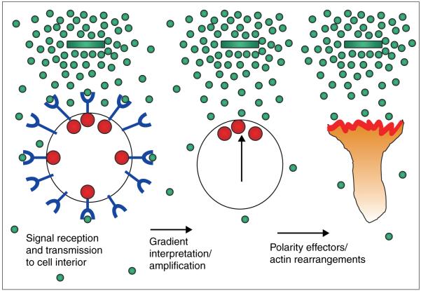
Requirements for eukaryotic chemotaxis. In order to respond appropriately to chemotactic gradients, eukaryotic cells must contain receptors (blue) that transmit a signal to the cell interior (red spheres) upon binding chemoattractant (green spheres). Each cell must manipulate this information to determine which region of its surface is exposed to maximal chemoattractant (vertical arrow). Finally, the cell must transmit this information to the final effectors responsible for spatial regulation of actin rearrangements and cell motility. Adapted from [26] and reproduced with permission.
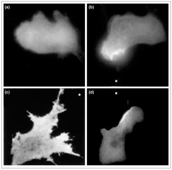
PI3K lipid products exhibit a polarized distribution during chemotaxis. The pleckstrin-homology domain of the protein kinase AKT/PKB fused to GFP was used as a probe for PI3K lipid products phosphatidylinositol 3,4-bisphosphate (PI[3,4]P2) and PIP3. (a) The probe is uniformly distributed throughout the cytosol of unstimulated neutrophil cells, but accumulates on the up-gradient face of cells exposed to chemoattractant. (b) Neutrophil-like HL60 cells exposed to a micropipette containing FMLP (asterisk). (c) 3T3 cell exposed to gradient of PDGF (asterisk). (d) Dictyostelium exposed to gradient of cAMP (asterisk). (a) and (b) are taken from [5••], (c) from [8••], and (d) was obtained courtesy of Satoru Funamoto and Rick Firtel.
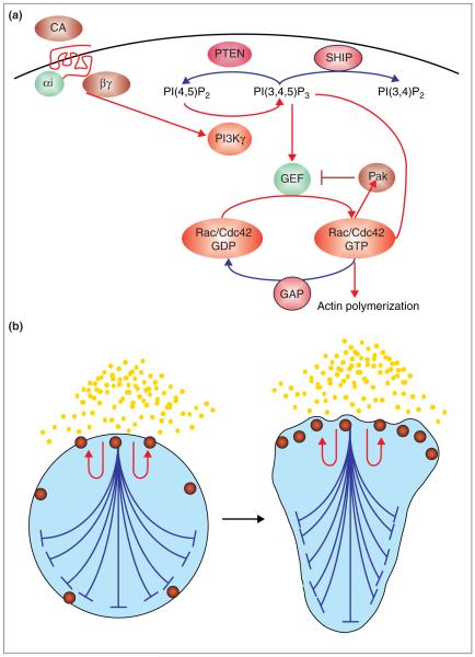
Signal processing that may orchestrate cell polarity during chemotaxis. (a) Summary of signal transduction cascade during chemotaxis. Red arrows denote positive regulators of the signal to chemotactic effectors, whereas the blue arrow denotes negative regulators of the signal. Binding of chemoattractant (CA) to G-protein-coupled chemoattractant receptors (red) results in the dissociation of G protein heterotrimer Gαi and Gβγ. Dissociated G protein activates PI3K (for neutrophils, directly through activation of PI3Kγ). PI3Kγ phosphorylates phosphatidylinositol 4,5-bisphosphate [PI{4,5}P2] to generate PIP3), which activates GEFs for the Rho GTPases Rac and Cdc42. The latter two proteins induce localized actin polymerization through Arp2/3 complex activation and other mechanisms. Two negative regulators of PIP3 accumulation are the phosphatases PTEN and SHIP, which generate PI(4,5)P2 and PI(3,4)P2, respectively. A negative-feedback loop for Cdc42 activation (from S. cerevisiae) involves a Pak-like serine/threonine kinase, which phosphorylates and inhibits the Cdc42 GEF, thereby decreasing the amount of active GTP-bound Cdc42. Finally, GTPase-activating proteins (GAPs) catalyse the conversion of active (GTP-bound) Rac/Cdc42 to inactive (GDP-bound) Rac/Cdc42. (b) Model for cell polarity during chemotaxis. A product of receptor activation (red spheres) increases its abundance in a short-range positive-feedback manner (red arrows) and also acts in a long-range inhibitory fashion to inhibit activation elsewhere (blue). Note that for simplicity the positive- and negative-feedback effects are only shown for one of the sites of activity. In reality, this process is occurring at sites of activity throughout the cell. The net effect of this pattern-formation system is to develop an amplified internal gradient of activity at the surface of the cell nearest the chemoattractant, or, in extreme cases, in response to stochastic differences in a uniform chemoattractant. Good candidates for the short-range positive-feedback loop are PIP3 , Rac and Cdc42. Good candidates for the long-range negative-feedback loop are phosphatases for PIP3 and negative regulation of the GEFs for Rac and Cdc42 (see [a]). For several recent mathematical models for chemotaxis, see [42-44].
References
-
-
Servant G, Weiner OD, Herzmark P, Balla T, Sedat JW, Bourne HR. Polarization of chemoattractant receptor signaling during neutrophil chemotaxis. Science. 2000;287:1037–1040.See annotation Haugh et al. (2000) [8••].
-
Publication types
MeSH terms
Substances
Grants and funding
LinkOut - more resources
Full Text Sources
Other Literature Sources
