. 2002 Mar;160(3):815-22.
doi: 10.1016/S0002-9440(10)64904-8.

Laser capture microdissection and two-dimensional polyacrylamide gel electrophoresis: evaluation of tissue preparation and sample limitations

Affiliations

Laser capture microdissection and two-dimensional polyacrylamide gel electrophoresis: evaluation of tissue preparation and sample limitations

Rachel A Craven et al. Am J Pathol. 2002 Mar.

Abstract

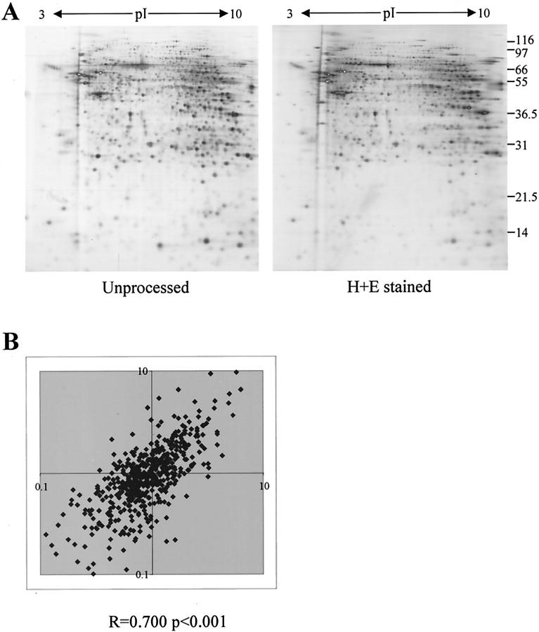
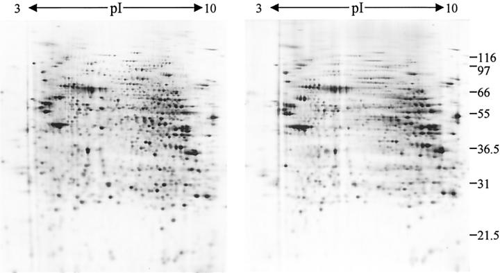
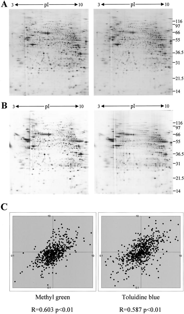
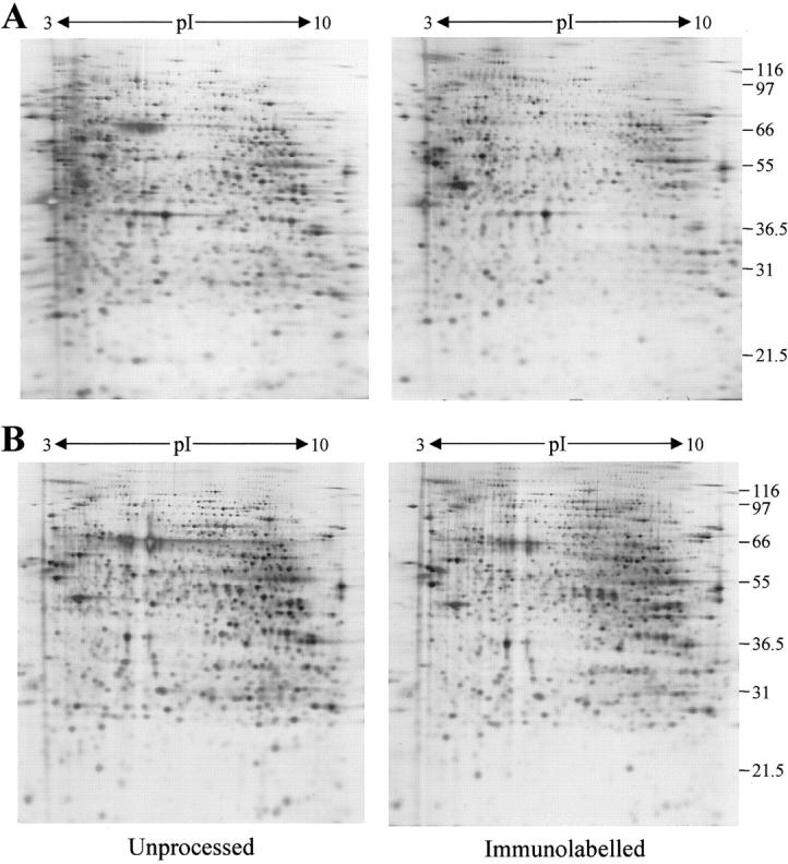
Laser capture microdissection (LCM) is now well established as a tool for facilitating the enrichment of cells of interest from tissue sections, overcoming the problem of tissue heterogeneity. LCM has been used extensively in combination with analysis at the DNA and RNA levels, but only a small number of studies have employed LCM with subsequent protein analysis, albeit with promising results. This study focuses on the potential of LCM in combination with two-dimensional polyacrylamide gel electrophoresis. The effects of tissue section preparation and sample type were evaluated to fully determine the suitability of using LCM in global protein profiling. The effects of several histochemical stains (hematoxylin and eosin, methyl green and toluidine blue) and immunolabeling on subsequent two-dimensional polyacrylamide gel electrophoresis were investigated. Quantitative analysis was performed to establish the extent of changes in the relative intensity of protein species and their reproducibility. All staining protocols tested were found to be compatible with protein analysis although there was variation in protein recovery and the quality of the protein profiles obtained. LCM of renal and cervix samples indicated that protein yield after dissection was acceptable, although the extent of enrichment and dissection time was tissue-dependent, which may preclude the use of this approach with some tissue types. These results indicate that LCM has potential as a tool in proteomic research.


Figures

Figure 1.
Figure 1.
The effect of H&E staining on 2D-gel profiles. A: 2D-gels of samples prepared from sections cut directly into lysis buffer (left) or onto slides and processed by H&E staining before protein extraction (right) are shown. B: The relative changes in intensity of 580 protein species in two independent processing experiments are shown. The ratio of percent volume of a spot in the control sample to percent volume of a spot in the H&E-stained sample was calculated after processing of two normal kidney cortex samples. The two ratios obtained for each spot were then plotted and a Spearman correlation coefficient was calculated for the data sets using SPSS for Windows.
Figure 2.
Figure 2.
The effect of eosin staining on 2D-PAGE. Two-dimensional gels of samples prepared from sections cut onto slides and stained with hematoxylin alone (left) or H&E (right) are shown. It should be noted that the effect of H&E staining on separation of proteins by 2D-PAGE was more pronounced in some experiments.
Figure 3.
Figure 3.
The effect of methyl green and toluidine blue staining on 2D-PAGE. Two-dimensional gels of samples prepared from sections cut directly into lysis buffer (left) or onto slides and processed (right) are shown for methyl green (A) and toluidine blue (B) staining. C: Scatter plots showing the conservation of relative change in intensity of protein species after processing are presented (for details see Figure 1 ▶ ).
Figure 4.
Figure 4.
Immunolabeling of proximal tubules in normal kidney cortex. Normal kidney cortex sections were labeled with anti-CD13 antibodies using a rapid-staining protocol that uses silver enhancement of gold labeling as the detection system.
Figure 5.
Figure 5.
Immunolabeling and 2D-gel analysis. Two-dimensional gels of samples prepared from sections of normal kidney cortex cut directly into lysis buffer (left) or onto slides and processed by immunolabeling before protein extraction (right) are shown. Immunolabeling was performed after acetone fixation (A) or ethanol fixation (B). Immunoglobulin heavy and light chains are not evident in the 2D-gel pattern, probably because the amount of bound antibody is below the detection limit.
Figure 6.
Figure 6.
LCM of normal cervical epithelium. Two-dimensional gels of dissected cervical epithelium (left), material remaining after dissection (stroma; right), and whole tissue sections (total; middle) are shown. A number of proteins with differential expression patterns are highlighted on the gels of stroma and epithelium. The numbered proteins are those for which identities have been determined (see text and Table 2 ▶ ). The gel load was normalized to the LCM sample that contained protein recovered from 7500 15-μm laser shots of epithelium from H&E-stained sections. Representative micrographs of the tissue samples are shown.

Similar articles

Cited by

References

    1. Page MJ, Amess B, Townsend RR, Parekh R, Herath A, Brusten L, Zvelebil MJ, Stein RC, Waterfield MD, Davies SC, O’Hare MJ: Proteomic definition of normal human luminal and myoepithelial breast cells purified from reduction mammoplasties. Proc Natl Acad Sci USA 1999, 96:12589-12594 - PMC - PubMed
    1. Reymond MA, Sanchez JC, Hughes GJ, Günther K, Riese J, Tortola S, Peinado MA, Kirchner T, Hohenberger W, Hochstrasser DF, Köckerling F: Standardized characterization of gene expression in human colorectal epithelium by two-dimensional electrophoresis. Electrophoresis 1997, 18:2842-2848 - PubMed
    1. Sarto C, Marocchi A, Sanchez JC, Giannone D, Frutiger S, Golaz O, Wilkins MR, Doro G, Cappellano F, Hughes G, Hochstrasser DF, Mocarelli P: Renal cell carcinoma and normal kidney protein expression. Electrophoresis 1997, 18:599-604 - PubMed
    1. Sirivatanauksorn Y, Drury R, Crnogorac-Jurcevic T, Sirivatanauksorn V, Lemoine NR: Laser-assisted microdissection: applications in molecular pathology. J Pathol 1999, 189:150-154 - PubMed
    1. Fend F, Raffeld M: Laser capture microdissection in pathology. J Clin Pathol 2000, 53:666-672 - PMC - PubMed

Publication types

MeSH terms