

Up-regulation of MHC class I expression accompanies but is not required for spontaneous myopathy in dysferlin-deficient SJL/J mice
- PMID: 11891182
- PMCID: PMC1867159
- DOI: 10.1016/S0002-9440(10)64906-1
Up-regulation of MHC class I expression accompanies but is not required for spontaneous myopathy in dysferlin-deficient SJL/J mice
Abstract
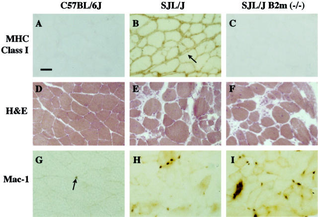
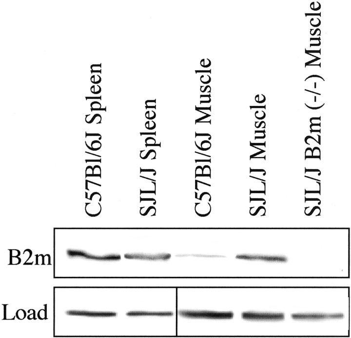
We found that up-regulation of major histocompatibility complex (MHC) class I expression accompanies, but is not required for, appearance of spontaneous myopathy in SJL/J mice. In some neuromuscular diseases, MHC class I expression is markedly up-regulated in muscles, though the consequences of this up-regulation for pathology are not clear. To study MHC class I in myopathy, we compared muscles of SJL/J mice to muscles of SJL/J mice that were also MHC class I-deficient due to targeted mutation in the beta-2-microglobulin gene (SJL/J B2m (-/-) mice). SJL/J mice show spontaneous myopathy and have a mutation in the dysferlin gene, a gene which is also mutated in human limb-girdle muscular dystrophy type 2B (LGMD2B). Muscles of eight-month-old SJL/J mice had higher levels of MHC class I expression than muscles of either C57BL/6J (wild-type) or SJL/J B2m (-/-) mice. In contrast, the percentage of abnormal muscle fibers was similar in SJL/J and SJL/J B2m (-/-) muscles. Invading Mac-1(+) cells were most abundant in SJL/J B2m (-/-) muscles, moderately abundant in SJL/J muscles, and rare in C57BL/6J muscles. Thus, MHC class I was markedly up-regulated in SJL/J muscles, but this high level of MHC class I was not necessary for the appearance of myopathy.
Figures
References
-
- Karpati G, Pouliot Y, Carpenter S: Expression of immunoreactive major histocompatibility complex products in human skeletal muscles. Ann Neurol 1988, 23:64-72 - PubMed
-
- Emslie-Smith AM, Arahata K, Engel AG: Major histocompatibility complex class I antigen expression, immunolocalization of interferon subtypes, and T cell-mediated cytotoxicity in myopathies. Hum Pathol 1989, 20:224-231 - PubMed
-
- McDouall RM, Dunn MJ, Dubowitz V: Expression of class I and class II MHC antigens in neuromuscular diseases. J Neurol Sci 1989, 89:213-226 - PubMed
-
- Nagaraju K, Raben N, Loeffler L, Parker T, Rochon PJ, Lee E, Danning C, Wada R, Thompson C, Bahtiyar G, Craft J, Hooft Van Huijsduijnen R, Plotz P: From the cover: conditional up-regulation of MHC class I in skeletal muscle leads to self-sustaining autoimmune myositis and myositis-specific autoantibodies. Proc Natl Acad Sci USA 2000, 97:9209–9214 - PMC - PubMed
-
- Hohlfeld R, Muller W, Toyka KV: Necrotizing myopathy in SJL mice. Muscle Nerve 1988, 11:184-185 - PubMed
Publication types
MeSH terms
Substances
LinkOut - more resources
Full Text Sources
Medical
Molecular Biology Databases
Research Materials
Miscellaneous
