. 2002 Mar;160(3):841-8.
doi: 10.1016/S0002-9440(10)64907-3.

CAP37, a novel inflammatory mediator: its expression in endothelial cells and localization to atherosclerotic lesions

Affiliations

CAP37, a novel inflammatory mediator: its expression in endothelial cells and localization to atherosclerotic lesions

Taunia D Lee et al. Am J Pathol. 2002 Mar.

Abstract

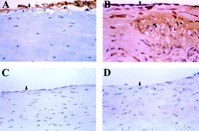
Cationic antimicrobial protein of 37 kd (CAP37), originally isolated from human neutrophils, is an important multifunctional inflammatory mediator. Here we describe its localization within the vascular endothelium associated with atherosclerotic plaques. Evidence from in vitro immunocytochemical, Northern blot, and reverse transcriptase-polymerase chain reaction analysis indicates that CAP37 is induced in endothelial cells in response to inflammatory mediators. Endothelial-derived CAP37 shows sequence identity with an extensive region of neutrophil-derived CAP37. This is the first demonstration of endogenous endothelial CAP37, confirmed by sequence analysis. We suggest that, because of its induction and location in the endothelium and its known monocyte- and endothelial-activating capabilities, CAP37 has potential to modulate monocyte/endothelial dynamics at the vessel wall in inflammation.


Figures

Figure 1.
Figure 1.
Localization of CAP37 in formalin-fixed, paraffin-embedded carotid artery A: Immunohistochemistry performed on atherosclerotic lesion present in the carotid artery using antisera to human CAP37 and the Vectastain Elite technique as described in the text. Strong staining (brown) indicated the presence of CAP37 in the endothelium. B: Detection of CAP37 in advanced atherosclerotic plaque indicating strong positive staining in endothelium and foam cells. C: Normal vessel stained with antisera to CAP37 indicating an absence of CAP37 in normal endothelium. D: Atherosclerotic lesion stained using an immunoadsorbed antisera to CAP37 that shows no staining. This lack of staining in D indicates the specificity of the antisera for CAP37 used in these assays. Sections were counterstained with hematoxylin. ↓, endothelium; *, foam cell. Original magnifications: ×400.
Figure 2.
Figure 2.
Induction of CAP37 protein in RAECs. A: Immunocytochemistry of RAECs stimulated with 10 μg/ml of LPS for 4 hours and stained with antisera to CAP37 using the Vectastain Elite ABC technique indicating strong staining (brown) for CAP37. B: RAECs incubated with media alone and stained with antisera to CAP37 shows no positive reaction, indicating that E-CAP37 is not constitutively expressed in RAECs. Sections counterstained with hematoxylin. Original magnifications: ×400 (A); ×200 (B).
Figure 3.
Figure 3.
Northern blot analysis of CAP37 mRNA in RAECs. Rat aorta endothelial cells were stimulated for 0 (lane 3), 0.5 (lane 4), 2 (lane 5), 4 (lane 6), 6 (lane 7), and 24 hours (lane 8) with S. minnesota LPS and the Northern blot performed on total RNA from each time point using the 32P-labeled CAP37 cDNA probe as described in text. An HL-60 cell line (lane 1) used as a positive control indicated presence of CAP37 mRNA (1000 bp). 18S and 28S rRNA (bottom) of total cellular RNA demonstrating the integrity and relative amounts of RNA. Lane 2 is empty.
Figure 4.
Figure 4.
RT-PCR analysis of HUVECs for CAP37 mRNA. Human umbilical vein endothelial cells were incubated with 10 ng/ml of TNF-α or left untreated (unt) for the indicated times and CAP37 mRNA expression (top, 468 bp) determined by RT-PCR. cDNA integrity was assessed with β-actin primers (bottom, 267 bp). This is a representative figure of five independent experiments.
Figure 5.
Figure 5.
Immunocytochemical assessment of surface-bound and cell-associated CAP37 in HUVECs. A: HUVECs incubated with TNF-α for 10 hours, fixed (but not permeabilized), and stained with antisera to CAP37 indicating no staining for CAP37 on the outer surface of the cell. B: Untreated HUVECs, fixed but not permeabilized and stained with antisera to CAP37 indicating lack of staining. C: HUVECs incubated with 10 ng/ml of TNF-α for 10 hours, permeabilized, and stained with antisera to CAP37 indicating strong cytoplasmic and perinuclear staining (brown) for CAP37. D: HUVECs incubated with media alone for 10 hours, permeabilized, and stained with antisera to CAP37 indicating light intracellular staining. E: HUVECs incubated with media alone for 10 hours, permeabilized, and stained with normal rabbit serum indicating no staining. Original magnifications: ×1000.
Figure 6.
Figure 6.
Flow cytometric analysis of cell-associated CAP37 in HUVECs. HUVECs were incubated 18 hours in the absence (unt) or presence of 10 ng/ml of TNF-α, permeabilized, and labeled with antisera to human CAP37 or normal serum control. Cells were permeabilized to determine intracellular levels of CAP37. A representative histogram from two independent experiments. The shift because of fluorescein isothiocyanate staining indicates increased expression of CAP37 in TNF-α stimulated cells. Also indicated is a low level of constitutive CAP37 expression (unt).
Figure 7.
Figure 7.
Western blot analysis of HUVECs for CAP37 protein. Human umbilical vein endothelial cells were incubated with TNF-α. Fifty μg of total protein was loaded into each lane. CAP37 protein expression, both cell-associated (lysate) and released (sup), was determined using rabbit antisera to human CAP37. PMN extract (20 μg) was included as a positive control for CAP37 staining.

References

    1. Shafer WM, Martin LE, Spitznagel JK: Cationic antimicrobial proteins isolated from human neutrophil granulocytes in the presence of diisopropyl fluorophosphate. Infect Immun 1984, 45:29-35 - PMC - PubMed
    1. Pereira HA, Spitznagel JK, Pohl J, Wilson DE, Morgan J, Palings I, Larrick JW: CAP37, a 37kD human neutrophil granule cationic protein shares homology with inflammatory proteinases. Life Sci 1990, 46:189-196 - PubMed
    1. Pohl J, Pereira HA, Martin NM, Spitznagel JK: Amino acid sequence of CAP37, a human neutrophil granule-derived antibacterial and monocyte-specific chemotactic glycoprotein structurally similar to neutrophil elastase. FEBS Lett 1990, 272:200-204 - PubMed
    1. Shafer WM, Martin LE, Spitznagel JK: Late intraphagosomal hydrogen ion concentration favors the in vitro antimicrobial capacity of a 37-kilodalton cationic granule protein of human neutrophil granules. Infect Immun 1986, 53:651-655 - PMC - PubMed
    1. Brackett DJ, Lerner MR, Lacquement MA, He R, Pereira HA: A synthetic lipopolysaccharide-binding peptide based on the neutrophil-derived protein CAP37 prevents endotoxin-induced responses in conscious rats. Infect Immun 1997, 65:2803-2811 - PMC - PubMed

Publication types

MeSH terms

LinkOut - more resources