. 2002 Mar;160(3):869-82.
doi: 10.1016/s0002-9440(10)64910-3.

Thymosin-beta4 regulates motility and metastasis of malignant mouse fibrosarcoma cells

Affiliations

Thymosin-beta4 regulates motility and metastasis of malignant mouse fibrosarcoma cells

Tokushige Kobayashi et al. Am J Pathol. 2002 Mar.

Abstract

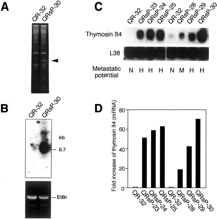
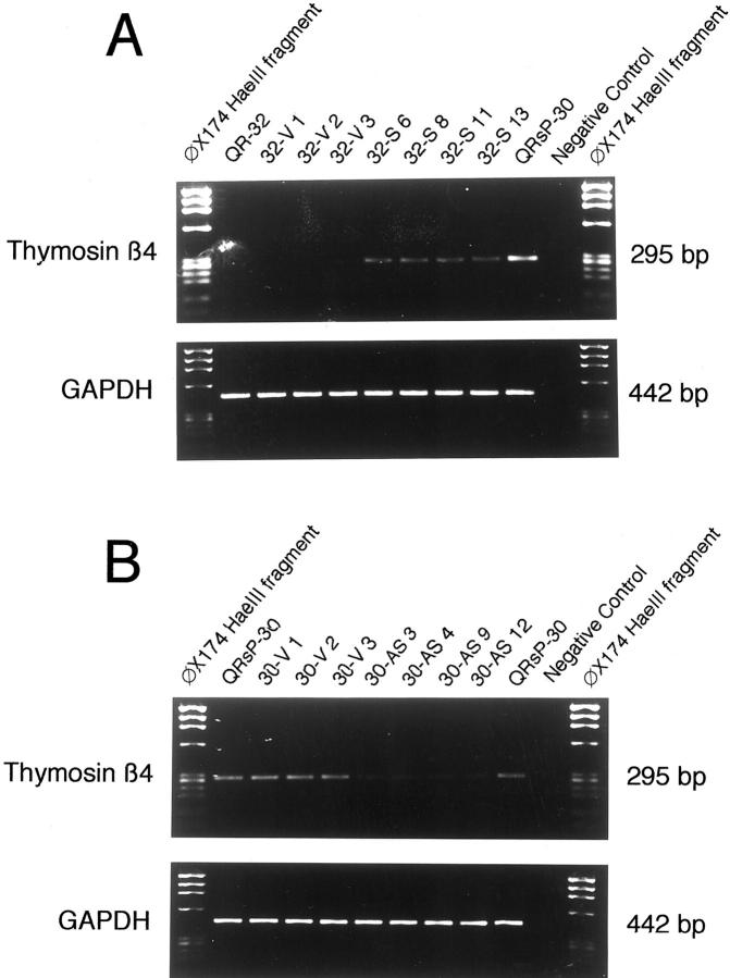
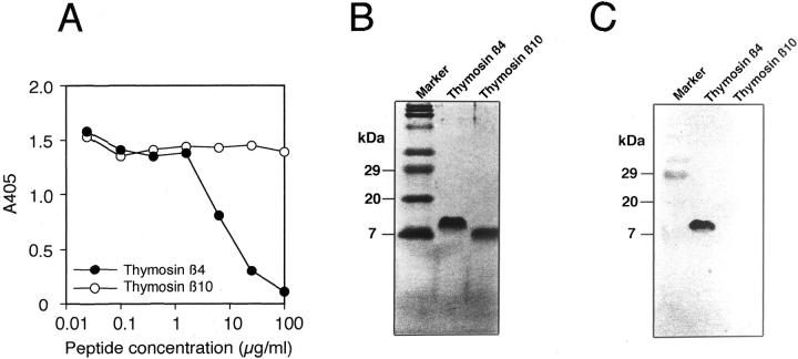
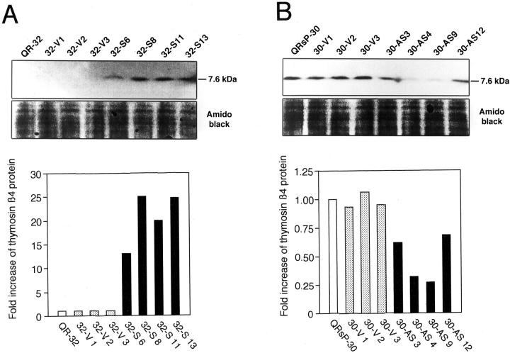
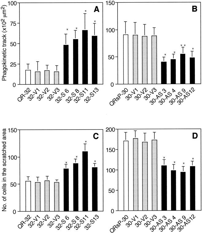
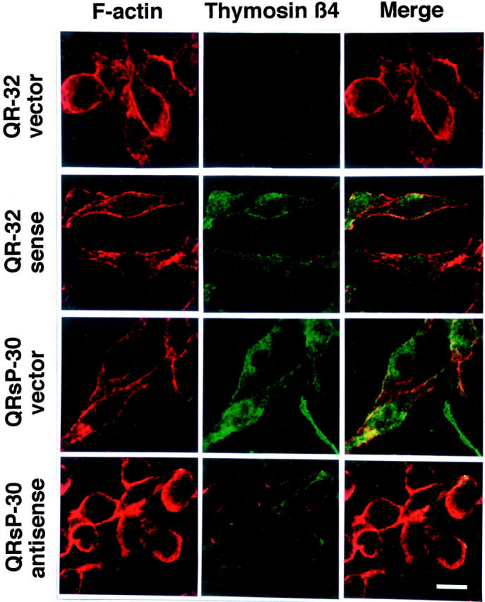
We identified a thymosin-beta4 gene overexpression in malignant mouse fibrosarcoma cells (QRsP-30) that were derived from clonal weakly tumorigenic and nonmetastatic QR-32 cells by using a differential display method. Thymosin-beta4 is known as a 4.9-kd polypeptide that interacts with G-actin and functions as a major actin-sequestering protein in cells. All of the six malignant fibrosarcoma cell lines that have been independently converted from QR-32 cells expressed high levels of thymosin-beta4 mRNA and its expression in tumor cells was correlated with tumorigenicity and metastatic potential. Up-regulation of thymosin-beta4 in QR-32 cells (32-S) transfected with sense thymosin-beta4 cDNA converted the cells to develop tumors and formed numerous lung metastases in syngeneic C57BL/6 mice. In contrast, antisense thymosin-beta4 cDNA-transfected QRsP-30 (30-AS) cells reduced thymosin-beta4 expression, and significantly lost tumor formation and metastases to distant organs. Vector-alone transfected cells (32-V or 30-V cells) behaved like their parental cells. We observed that tumor cell motility, cell shape, and F-actin organization is regulated in proportion to the level of thymosin-beta4 expression. These findings indicate that thymosin-beta4 molecule regulates fibrosarcoma cell tumorigenicity and metastasis through actin-based cytoskeletal organization.


Figures

Figure 1.
Figure 1.
Differential display and Northern blot reveal increased expression of thymosin-β4 in malignant mouse fibrosarcoma cell lines as compared to parental QR-32 cells. A: Differential display demonstrates markedly augmented expression of thymosin-β4 (arrowhead) in malignant fibrosarcoma cells, QRsP-30 when compared with the parental QR-32 cells. B: Northern blot using thymosin-β4 gene as probe identifies a 0.7-kb mRNA expression in QRsP-30 cells (top). 28S rRNA expression served as a loading control (bottom). C: Northern blot confirmed that the expression of thymosin-β4 gene was enhanced in malignant tumor cell lines. L38 hybridization served as a loading control. Metastatic potential of the intravenously injected tumor cells (as described in Materials and Methods). N, nonmetastatic; M, moderately metastatic (forming metastatic colonies not exceeding 150 per lung); H, highly metastatic (forming colonies more than 150 per lung). D: The expression of the thymosin-β4 mRNA comparing L38 mRNA were quantified by densitometry.
Figure 2.
Figure 2.
RT-PCR analysis of thymosin-β4 expression in the cells transfected with sense or antisense thymosin-β4 construct and vector controls. RT-PCR analysis of parent cells and its transfectants. Thymosin-β4 at 295 bp in sense thymosin-β4 transfectants was increased, whereas it was reduced in antisense thymosin-β4 transfectants. A: QR-32 cells transfected with sense thymosin-β4 construct. B: QRsP-30 cells transfected with antisense thymosin-β4 construct.
Figure 3.
Figure 3.
Specificity of the monoclonal antibody, TB4N1-5, as determined by Western blot and competitive inhibition assay. A: Peptide competitive inhibition assay. The 96-well plates were precoated with synthetic peptide of thymosin-β4 (2 μg/ml). TB4N1-5 antibody (5 μg/ml) was added to the wells with serially diluted synthetic peptide, thymosin-β4 (•) or thymosin-β10 (○). Absorbance at 405 nm was measured after incubation with ABTS and HRP-conjugated anti-mouse immunoglobulins as described in Materials and Methods. B: Coomassie brilliant blue staining. The synthetic peptides of thymosin-β4 and thymosin-β10 (1.5 μg/lane) were electrophoresed in SDS-polyacrylamide gel electrophoresis and the gel was stained with Coomassie brilliant blue solution. C: Western blotting by TB4N1-5 antibody. Another gel, electrophoresed as the same procedures as described in A, were transferred onto membrane and reacted with TB4N1-5 antibody (1 μg/ml).
Figure 4.
Figure 4.
Western blot of thymosin-β4 protein levels in the cells transfected with sense or antisense thymosin-β4 construct and vector controls. Western blot analysis of parent cells and its transfectants. Thymosin-β4 (Mr 7600) in sense thymosin-β4 transfectants was increased, whereas it was reduced in antisense thymosin-β4 transfectants. The expression of thymosin-β4 proteins comparing Amido Black staining were quantified by densitometry. A: QR-32 cells transfected with sense thymosin-β4 construct. B: QRsP-30 cells transfected with antisense thymosin-β4 construct.
Figure 5.
Figure 5.
Phagokinetic track patterns produced by sense (A) or antisense (B) thymosin-β4 transfectants and their control tumor cells. The gold particles were scattered on the glass coverslips. Tumor cells were plated on the glass coverslips in culture. After 48 hours of incubation, the phagokinetic track areas of 40 individual cells were measured and the average area is shown. Each bar represents the means ± SD of three independent experiments done in duplicate. *, P < 0.001; **, P < 0.005, compared with an empty-vector transfectant. Scratch-wound closure patterns produced by sense (C) or antisense (D) thymosin-β4 transfectants and their control tumor cells. An in vitro wound was introduced in confluent cultures of each cell lines. After 12 hours, the cells were photographed. The number of cells migrated into the scratched area was counted. Each bar represents the means ± SD of three independent experiments done in duplicate. *, P < 0.001, compared with an empty-vector transfectant.
Figure 6.
Figure 6.
Thymosin-β4 levels inversely correlates with F-actin staining. Benign or malignant mouse fibrosarcoma cells expressing vector construct, and sense and antisense thymosin-β4 cDNA has been attached onto glass coverslips, then fixed and stained with thymosin-β4 antibody (green) and Texas Red-X phalloidin (red) to detect thymosin-β4 distribution and F-actin, respectively, and observed by confocal scanning laser microscopy. Original magnification of each clone, ×630. Representative image of three independent experiments with similar results is shown. Scale bar, 10 μm.

Similar articles

Cited by

References

    1. Steeg PS, Bevilacqua G, Kopper L, Thorgeirsson UP, Talmadge JE, Liotta LA, Sobel ME: Evidence for a novel gene associated with low tumor metastatic potential. J Natl Cancer Inst 1988, 80:200-204 - PubMed
    1. Lee J-H, Miele ME, Hicks DJ, Phillips KK, Trent JM, Weissman BE, Welch DR: KiSS-1, a novel human malignant melanoma metastasis-suppressor gene. J Natl Cancer Inst 1996, 88:1731-1737 - PubMed
    1. Ebralidze A, Tulchinsky E, Grigorian M, Afanasyeva A, Senin V, Revazova E, Lukanidin E: Isolation and characterization of a gene specifically expressed in different metastatic cells and whose deduced gene product has a high degree of homology to a Ca2+-binding protein family. Genes Dev 1989, 3:1086-1093 - PubMed
    1. Gunthert U, Hofmann M, Rudy W, Reber S, Zoller M, Haussmann I, Matzku S, Wenzel A, Ponta H, Herrlich P: A new variant of glycoprotein CD44 confers metastatic potential to rat carcinoma cells. Cell 1991, 65:13-24 - PubMed
    1. Okada F, Hosokawa M, Hamada J-I, Hasegawa J, Koto M, Mizutani T, Ren J, Takeichi N, Kobayashi H: Malignant progression of a mouse fibrosarcoma by host cells reactive to a foreign body (gelatin sponge). Br J Cancer 1992, 66:635-639 - PMC - PubMed

Publication types