

In situ analysis of the variable heavy chain gene of an IgM/IgG-expressing follicular lymphoma: evidence for interfollicular trafficking of tumor cells
- PMID: 11891187
- PMCID: PMC1867185
- DOI: 10.1016/S0002-9440(10)64911-5
In situ analysis of the variable heavy chain gene of an IgM/IgG-expressing follicular lymphoma: evidence for interfollicular trafficking of tumor cells
Abstract
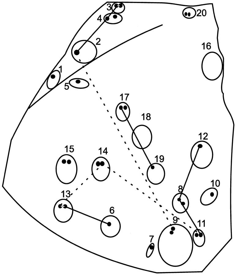
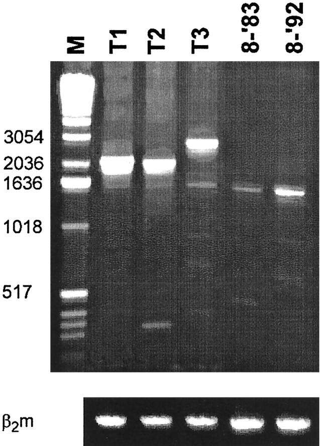
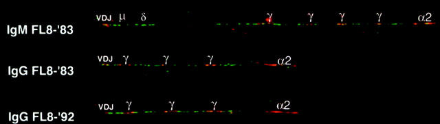
It is generally assumed that follicular lymphomas (FL) not only morphologically resemble normal germinal centers but have retained some functional characteristics of their non-neoplastic counterparts as well. Recent IgV gene analyses on a panel of FLs however, strongly suggested that FLs do not retain the capacity of somatic hypermutation and are not being selected on basis of the quality of their mIgV regions. To extend these findings, we investigated the follicular organization and class switching in a FL that consisted of both IgM- and IgG-expressing tumor cells with a high somatic mutation load and significant intraclonal V(H) gene diversity. V(H)-C(mu) and V(H)-Cgamma gene transcripts were amplified and sequenced from samples of approximately 50 tumor cells, isolated from frozen tissue sections by laser microdissection. We identified many different subclones and obtained limited evidence of subclone dominance in individual follicles. Remarkably, several subclones were found scattered over different follicles. All samples contained IgM- and IgG-expressing tumor cells with, in general, non-identical mutation patterns, which is not in support of ongoing class switching. Accordingly, no switch circle recombination products were found. The findings indicate that the neoplastic follicles lack the organization and functions typical of reactive germinal centers.
Figures
Similar articles
-
Variable heavy chain gene analysis of follicular lymphomas: correlation between heavy chain isotype expression and somatic mutation load.Blood. 2000 May 1;95(9):2922-9. Blood. 2000. PMID: 10779440
-
Variable heavy-chain gene analysis of follicular lymphomas: subclone selection rather than clonal evolution over time.Blood. 2001 Jul 1;98(1):238-40. doi: 10.1182/blood.v98.1.238. Blood. 2001. PMID: 11418487
-
IgM-producing chronic lymphocytic leukemia cells undergo immunoglobulin isotype-switching without acquiring somatic mutations.J Clin Invest. 1996 Jul 15;98(2):290-8. doi: 10.1172/JCI118792. J Clin Invest. 1996. PMID: 8755637 Free PMC article.
-
Immunoglobulin receptor evolution in follicular lymphoma and a review of literature.Leuk Lymphoma. 2007 Oct;48(10):2063-7. doi: 10.1080/10428190701540983. Leuk Lymphoma. 2007. PMID: 17917975 Review. No abstract available.
-
Hypermutation in antibody affinity maturation.Curr Opin Immunol. 1999 Apr;11(2):186-9. doi: 10.1016/s0952-7915(99)80031-4. Curr Opin Immunol. 1999. PMID: 10322147 Review.
Cited by
-
Pathogenesis of follicular lymphoma.J Clin Invest. 2012 Oct;122(10):3424-31. doi: 10.1172/JCI63186. Epub 2012 Oct 1. J Clin Invest. 2012. PMID: 23023713 Free PMC article. Review.
-
Somatic hypermutation analysis in follicular lymphoma provides evidence suggesting bidirectional cell migration between lymph node and bone marrow during disease progression and relapse.Haematologica. 2013 Sep;98(9):1433-41. doi: 10.3324/haematol.2012.074252. Epub 2013 Apr 12. Haematologica. 2013. PMID: 23585531 Free PMC article.
References
-
- Rajewsky K: Clonal selection and learning in the antibody system. Nature 1996, 381:751-758 - PubMed
-
- Lam K-P, Kühn R, Rajewsky K: In vivo ablation of surface immunoglobulin on mature B cells by inducible gene targeting results in rapid cell death. Cell 1997, 90:1073-1083 - PubMed
-
- Lindhout E, Koopman G, Pals ST, de Groot C: Triple check for antigen specificity of B cells during germinal center reactions. Immunol Today 1997, 18:573-577 - PubMed
-
- Kocks C, Rajewsky K: Stable expression and somatic hypermutation of antibody V regions in B cell developmental pathways. Annu Rev Immunol 1989, 7:537-559 - PubMed
Publication types
MeSH terms
Substances
LinkOut - more resources
Full Text Sources
Research Materials
