

Genetic and immunohistochemical analysis of pancreatic acinar cell carcinoma: frequent allelic loss on chromosome 11p and alterations in the APC/beta-catenin pathway
- PMID: 11891193
- PMCID: PMC1867188
- DOI: 10.1016/s0002-9440(10)64917-6
Genetic and immunohistochemical analysis of pancreatic acinar cell carcinoma: frequent allelic loss on chromosome 11p and alterations in the APC/beta-catenin pathway
Abstract
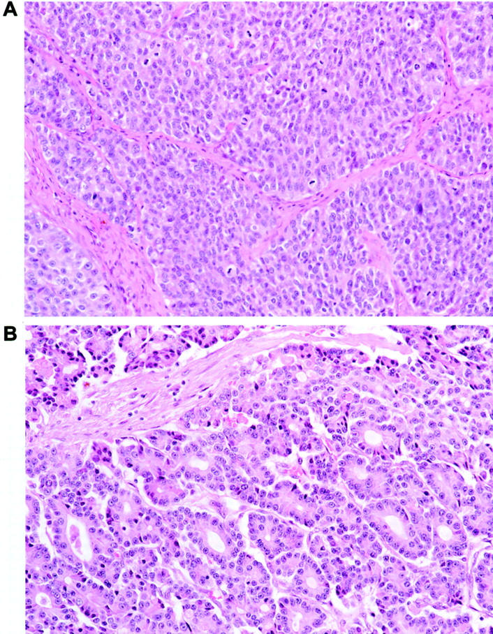
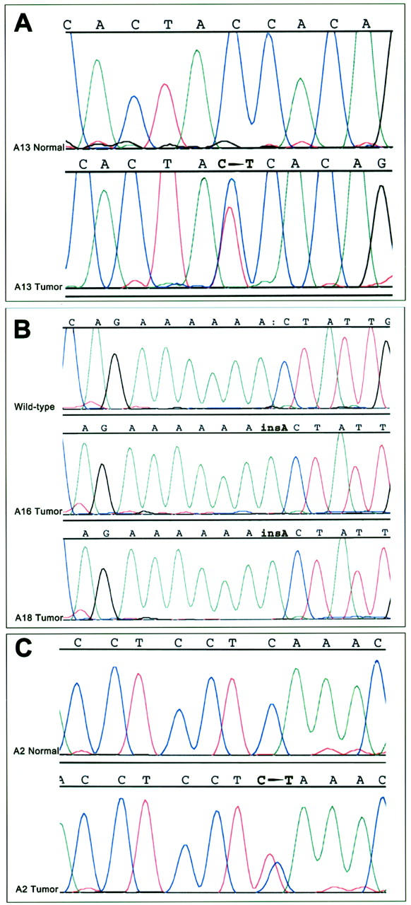
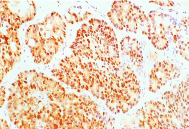
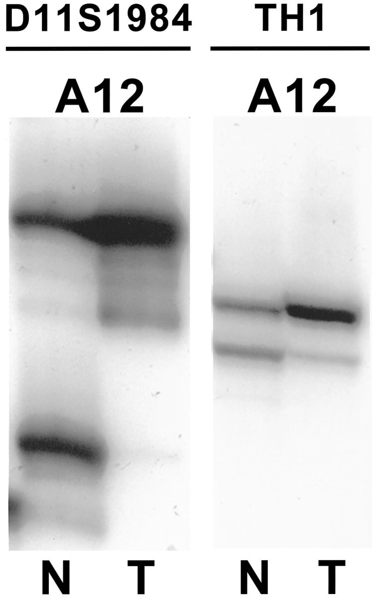
Acinar cell carcinomas (ACCs) are rare malignant tumors of the exocrine pancreas. The specific molecular alterations that characterize ACCs have not yet been elucidated. ACCs are morphologically and genetically distinct from the more common pancreatic ductal adenocarcinomas. Instead, the morphological, immunohistochemical, and clinical features of ACCs overlap with those of another rare pancreatic neoplasm, pancreatoblastoma. We have recently demonstrated a high frequency of allelic loss on chromosome arm 11p and mutations in the APC/beta-catenin pathway in pancreatoblastomas, suggesting that similar alterations might also play a role in the pathogenesis of some ACCs. We analyzed a series of 21 ACCs for somatic alterations in the APC/beta-catenin pathway and for allelic loss on chromosome 11p. In addition, we evaluated the ACCs for alterations in p53 and Dpc4 expression using immunohistochemistry, and for microsatellite instability (MSI) using polymerase chain amplification of a panel of microsatellite markers. Allelic loss on chromosome 11p was the most common genetic alteration in ACCs, present in 50% (6 of 12 informative cases). Molecular alterations in the APC/beta-catenin pathway were detected in 23.5% (4 of 17) of the carcinomas, including one ACC with an activating mutation of the beta-catenin oncogene and three ACCs with truncating APC mutations. One ACC (1 of 13, 7.6%) showed allelic shifts in four of the five markers tested (MSI-high), two (15.4%) showed an allelic shift in only one of the five markers tested (MSI-low), and no shifts were detected in the remaining 10 cases. The MSI-high ACC showed medullary histological features. In contrast, no loss of Dpc4 protein expression or p53 accumulation was detected. These results indicate that ACCs are genetically distinct from pancreatic ductal adenocarcinomas, but some cases contain genetic alterations common to histologically similar pancreatoblastomas.
Figures

References
-
- Klimstra DS, Heffess CS, Oertel JE, Rosai J: Acinar cell carcinoma of the pancreas. A clinicopathologic study of 28 cases. Am J Surg Pathol 1992, 16:815–837 - PubMed
-
- Chen J, Baithun SI, Ramsay MA: Histogenesis of pancreatic carcinomas: a study based on 248 cases. J Pathol 1985, 146:65-76 - PubMed
-
- Morohoshi T, Kanda M, Horie A, Chott A, Dreyer T, Kloppel G, Heitz PU: Immunocytochemical markers of uncommon pancreatic tumors. Acinar cell carcinoma, pancreatoblastoma, and solid cystic (papillary-cystic) tumor. Cancer 1987, 59:739-747 - PubMed
-
- Shinagawa T, Tadokoro M, Maeyama S, Maeda C, Yamaguchi S, Morohoshi T, Ishikawa E: Alpha fetoprotein-producing acinar cell carcinoma of the pancreas showing multiple lines of differentiation. Virchows Arch 1995, 426:419-423 - PubMed
-
- Caruso RA, Inferrera A, Tuccari G, Barresi G: Acinar cell carcinoma of the pancreas. A histologic, immunocytochemical and ultrastructural study. Histol Histopathol 1994, 9:53-58 - PubMed
Publication types
MeSH terms
Substances
Grants and funding
LinkOut - more resources
Full Text Sources
Other Literature Sources
Medical
Molecular Biology Databases
Research Materials
Miscellaneous
