. 2002 Mar;160(3):1021-34.
doi: 10.1016/S0002-9440(10)64923-1.

Fibrinogen stabilizes placental-maternal attachment during embryonic development in the mouse

Affiliations

Fibrinogen stabilizes placental-maternal attachment during embryonic development in the mouse

Takayuki Iwaki et al. Am J Pathol. 2002 Mar.

Abstract

In humans, maternal fibrinogen (Fg) is required to support pregnancies by maintaining hemostatic balance and stabilizing uteroplacental attachment at the fibrinoid layer found at the fetal-maternal junction. To examine relationships between low Fg levels and early fetal loss, a genetic model of afibrinogenemia was developed. Pregnant mice homozygous for a deletion of the Fg-gamma chain, which results in a total Fg deficiency state (FG(-/-)), aborted the fetuses at the equivalent gestational stage seen in humans. Results obtained from timed matings of FG(-/-) mice showed that vaginal bleeding was initiated as early as embryonic day (E)6 to 7, a critical stage for maternal-fetal vascular development. The condition of afibrinogenemia retarded embryo-placental development, and consistently led to abortion and maternal death at E9.75. Lack of Fg did not alter the extent or distribution pattern of other putative factors of embryo-placental attachment, including laminin, fibronectin, and Factor XIII, indicating that the presence of fibrin(ogen) is required to confer sufficient stability at the placental-decidual interface. The results of these studies demonstrate that maternal Fg plays a critical role in maintenance of pregnancy in mice, both by supporting proper development of fetal-maternal vascular communication and stabilization of embryo implantation.


Figures

Figure 1.
Figure 1.
Murine E6.0 uterus and ovaries (original magnifications, ×100). Morphological characterization (H&E staining) of the uteri and ovaries of WT and Fg-deficient (FG−/−) mice on day 6 of pregnancy (E6). In both WT (A) and FG−/− (B) pregnant mice, implanted embryos appear normal. In both embryos, the ectoplacental cone (EC) seems to be at the same developmental stage. Maternal bleeding (asterisk) is seen at the implantation site in the uterus of the FG−/− mouse (B). In ovaries of both genotypes (C and D), evidence of luteinization and intact corpus luteum is seen, along with healthy follicles containing ova (arrows).
Figure 2.
Figure 2.
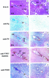
The murine E7.0 placenta (original magnifications, ×100). A and B: H&E staining of the WT egg cylinder containing an embryo. Mild bleeding is observed in the ectoplacental cone (EC) region (A) that is more pronounced in a similarly stained section of a FG−/− female (B, unlabeled arrows). Bleeding is seen in the egg cylinder cavity of the FG−/− mouse and is also observed in the anti-mesometrial decidual (DE) areas in A and B. The primitive streak (PS) is also indicated in A. C: Anti-Fg/fibrin immunostaining shows Fg (or fibrin) (reddish-brown) at the sites of the blood pools in a WT mouse. This is obviously not present in a FG−/− mouse (D). Fg/fibrin staining is also located around the embryonic endoderm in C, as well as at the EC, the implantation site (IS), DE, and endometrium (EM). E and F: Anti-FN immunostaining shows that this protein is also located throughout the EC, as well as at the embryonic region distal to the IS region and the IS itself, demonstrating that trabecular processes are occurring. Some staining occurs in the DE that is pronounced at the IS. FN is heavily stained throughout the endometrium in both WT (E) and FG−/− (F) mice. G and H: Double immunostaining of VWF (brown) and laminin (gray) on WT (G) and FG−/− (H) mice tissues. The anti-VWF staining shows numerous endothelial cell-lined vessels in the DE, the EC, the embryonic distal areas, and at the IS of both genotypes. Laminin is also present throughout the DE and EM. The amniotic (AC) and ectoplacental cavities (EPC) are indicated. I and J: Anti-FXIII immunostaining of WT (I) and FG−/− (J) tissue sections reveals the presence of this protein lining outer membrane regions of the embryos. FXIII appears to be localized around Fg and FN in the implantation site in the WT mouse (I), and with FN in the FG−/− mouse (J).
Figure 3.
Figure 3.
The murine E8.0 placenta (original magnifications, ×100). These embryos were whole-mount stained with X-Gal before sectioning. Embryonic tissue is thus stained a bluish-green. In some cases, eg, H&E staining, this embryonic coloration is not visible because it is overtaken by other stains. A and B: H&E stainings of WT and FG−/− pregnant dams, emphasizing the ectoplacental cone and implantation site regions in the developing chorioallantoic placenta, show blood-containing sinuses and vessels in these regions in both genotypes, with some associated bleeding events outside of the yolk sac cavity that occur in both WT and FG−/− mice. C: The implantation site, as well as regions of the decidua, stained positively for Fg/fibrin (reddish-brown) in the WT mouse, likely from maternal vessels, but obviously not in the FG−/− animal (D). E and F: FN immunostaining (reddish-brown) is co-localized with Fg in the WT mouse placenta (E) and is present at the implantation site and in stromal regions of the decidua in the FG−/− mouse placenta (F). The embryo also contains some FN staining at this gestational age. G and H: VWF immunostaining (reddish-brown) indicates the presence of endothelial cell-lined vessels in the ectoplacental cone and implantation site regions of both genotypes. These vessels are the likely vehicle of transit of maternal Fg and FXIII found at these sites. The increased levels of VWF at the fibrinoid layer in the FG−/− mouse (H) probably arise from platelets that aggregate to attempt to stop bleeding. Laminin (gray, but slightly discolored by the counterstain) stains heavily throughout the decidua and embryo. I and J: Anti-FXIII immunostaining (reddish-brown) showed the presence of this protein co-localized with Fg and FN in the WT mouse, and with FN in the FG−/− mouse. Staining is also observed in stromal regions of the decidua.
Figure 4.
Figure 4.
The murine E.9.0 placenta (original magnifications, ×200). A and B: H&E staining of WT and FG−/− embryos shows the attachment of the labyrinth (L) to spongiotrophoblasts (ST) in the WT tissue (A). The giant cell (GC) layer is also observable at the interface between the decidua (DE) and ST layers. B: The placenta in the FG−/− mouse appears smaller than its WT counterpart, and the ST layers are beginning to separate from the DE. C and D: Anti-Fg/fibrin (reddish-brown) immunostaining clearly shows a Fg/fibrin-rich fibrinoid layer between the GC and ST layers (C). None of this staining is seen the FG−/− mouse tissue (D). E and F: Anti-FN staining (gray-black) is co-localized with Fg/fibrin in the fibrinoid layer in the WT (E) and FG−/− (F) placentas. G and H: Anti-VWF staining (reddish-brown) demonstrates the presence of endothelial cell-lined vessels in the fibrinoid layer and DE of both the WT (G) and FG−/− (H) tissue sections, that are likely supplying the maternal circulation to the embryo. Very little staining is observed in the labyrinth, suggesting that the large sinuses present therein are not derived from the endothelium. Anti-laminin staining (gray) of the same tissue shows the presence of laminin throughout the ST, L, and DE layers in both genotypes. I and J: Anti-FXIII immunostaining (reddish-brown) demonstrates the presence of FXIII in the interfacial fibrinoid layer of both genotypes, as well in stromal regions of the DE.
Figure 5.
Figure 5.
The murine WT E9.75 placenta (original magnifications, ×100). A: H&E staining of WT sections illustrating the fusion of the maternal decidua (DE) with the embryonic layers in the WT placenta. The labyrinth (L) layer, which is fused to the spongiotrophoblast (ST) layer, followed into the DE by the giant cell (GC) layer, is clearly observed, as is the chorionic plate (CP). B: Anti-Fg/fibrin immunostaining (reddish-brown) of serial sections of this tissue indicates the presence of the Fg/fibrin-containing fibrinoid layer (FL), and staining throughout the DE. C: Anti-FN immunostaining (gray/black) indicates the co-localization of this protein with Fg/fibrin in the FL. D: Anti-VWF immunostaining (reddish-brown) demonstrates that endothelial cell-lined vessels are present in the maternal and embryonic regions of the placenta. Anti-VWF staining is also co-localized with that of Fg/fibrin and FN. Laminin (gray) is present throughout the labyrinth and decidual regions. E: Anti-FXIII immunostaining (reddish-brown) shows the presence of this protein in the FL and its co-localization with Fg/fibrin and FN.
Figure 6.
Figure 6.
The murine E9.75 egg cylinders of pregnant WT and FG−/− mice (original magnifications, ×40). These samples were whole-mount stained with X-Gal before sectioning. A: H&E staining of the E9.75 WT mouse egg cylinder shows a normally developing embryo with its yolk sac (YS) lining the cylinder wall. YS vasculature and blood lakes are seen (black arrows). The uterine implantation site (IS) in the decidua (DE) is indicated. H&E (B) and anti-Fg/fibrin (C) stainings of the FG−/− E9.75 egg cylinder illustrate a massive bleeding event (asterisk) in the egg cylinder cavity. The presence of embryonic cells (filled arrow) in a DE site that is remote from the position of the fetus (C) indicates that placental detachment likely has occurred from this site (the implantation site). An expanded view of the area in the vicinity of the black arrow shows the presence of embryonic giant cells (red arrows) in this same region. E: Anti-FN staining shows the presence of FN in the same region of the DE (filled arrow) as the embryonic cells in C. GC (red arrows, inset) associated with the FN layer are clearly seen in an expanded view (F) of a region of the black arrow in E. This further suggests that this area was the original implantation site. G: Anti-VWF/anti-laminin and FXIII stainings show numerous endothelial cell-lined vessels and perhaps platelets (arrow), which is observed more clearly in an expanded view of the implantation site (H). I: Anti-FXIII immunostaining demonstrates co-localization of this protein with Fg/fibrin, FN, and VWF at the implantation site. J: An expanded view of the arrowed region of I shows embryonic giant cells (red arrows) around the FXIII immunostains. Original magnifications, ×40 (A–C, E, G, and I); D, F, H, and J were arbitrarily expanded for purposes of ease of visualization.
Figure 7.
Figure 7.
Plasma levels of Fg during pregnancies of various genotypes. A: The consumption of exogenous Fg by pregnant FG−/− mice. A quantity of 10 mg of Fg was injected into pregnant females at E4.5 and blood drawn for 3 consecutive days. Other injections of the same quantity were made every second day (arrows) and Fg levels determined. The dotted vertical line represents the lowest level of Fg detection in our assay. B: The Fg concentration in plasmas of WT (black bars) and heterozygous (FG+/−) pregnant mice (gray bars) during the course of pregnancy. Plasma was taken by eye bleedings from individual WT mice (n = 8) and FG+/− mice (n = 14 to 16). Fg levels were determined at E3.5, E8.5, E13.5, and E18.5.
Figure 8.
Figure 8.
Rescue of pregnancies in E.9.75 FG−/− pregnant mice as a result of Fg administration. Gross examinations of E9.75 uteri containing embryos within their egg cylinders show severe intrauterine bleeding in the FG−/− mouse (A), as compared to a WT mouse (B). C: The egg cylinders of E9.75 FG−/− mouse that was administered Fg possessed the gross features as the WT embryos and egg cylinders. D: H&E stain (original magnification, ×100) of the placenta and implantation site (IS) of a rescued E9.75 FG−/− mouse embryo shows a normal labyrinth (L) layer, with embryonic blood in large sinuses attached to the yolk sac (YS). A spongiotrophoblast (ST)-containing area is present, with an interfacial layer of giant cells (GC) between the ST and decidual (DE) regions. A large blood-filled maternal artery is observed in the DE. E: Anti-Fg/fibrin (reddish-brown) staining (original magnification, ×100) of a serial section of D of the tissue of the treated FG−/− mouse, showing that Fg that was administered to plasma of the FG−/− mother is present in vessels within the DE, as well as in the fibrinoid layer (FL) at the IS (asterisk). Randomized staining is also present in the vessels in the embryonic (blue-green) ST area, but is not visible in the vessels of the labyrinthine region. F: Anti-FN (reddish-brown) staining (original magnification, ×100) shows the presence of FN surrounding macrophages in the DE layer and some ST cells of the Fg-treated FG−/− mother. This protein is abundant in the labyrinth, especially around the large sinuses (SN), as well as in the chorionic plate, and co-localizes with Fg/fibrin at the FL at the IS (asterisk). G: Anti-VWF (reddish-brown)/anti-laminin (gray) staining (original magnification, ×100) illustrates the endothelial cell-lined vessels of the maternal DE, and the embryonic ST, labyrinth, and YS areas of the treated E9.75 FG−/− mouse. H: Anti-FXIII (reddish-brown) staining (original magnification, ×100) shows abundant FXIII immunostaining in the DE area, including the FL. I: A ×40 view of the anti-FN (reddish-brown)-stained tissue section (F). FN staining in the treated FG−/− mouse is also noted, lining the dorsal aorta of the embryo (asterisk), as well as the amnion (AM). The now well-vascularized YS fully lines the cylinder cavity, and is clearly attached to the IS. Embryos used in D to H were whole-mount stained with X-Gal before sectioning.

Similar articles

Cited by

References

    1. Kant JA, Fornace Jr AJ, Saxe D, Simon MI, McBride OW, Crabtree GR: Evolution and organization of the fibrinogen locus on chromosome 4: gene duplication accompanied by transposition and inversion. Proc Natl Acad Sci USA 1985, 82:2344–2348 - PMC - PubMed
    1. Doolittle RF: Fibrinogen and fibrin. Annu Rev Biochem 1984, 53:195-229 - PubMed
    1. McRitchie DI, Girotti MJ, Glynn MF, Goldberg JM, Rotstein OD: Effect of systemic fibrinogen depletion on intraabdominal abscess formation. J Lab Clin Med 1991, 118:48-55 - PubMed
    1. Kuijper PH, Gallardo-Torres HI, Lammers JW, Sixma JJ, Koenderman L, Zwaginga JJ: Platelet and fibrin deposition at the damaged vessel wall: cooperative substrates for neutrophil adhesion under flow conditions. Blood 1997, 89:166-175 - PubMed
    1. Martin P: Wound healing—aiming for perfect skin regeneration. Science 1997, 276:75-81 - PubMed

Publication types

MeSH terms