. 2002 May 24;277(21):19056-63.
doi: 10.1074/jbc.M110960200. Epub 2002 Mar 12.

Identification by site-directed mutagenesis of residues involved in ligand recognition and activation of the human A3 adenosine receptor

Affiliations

Identification by site-directed mutagenesis of residues involved in ligand recognition and activation of the human A3 adenosine receptor

Zhan-Guo Gao et al. J Biol Chem. .

Abstract

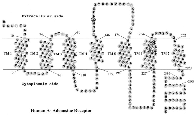
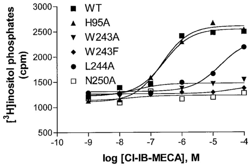
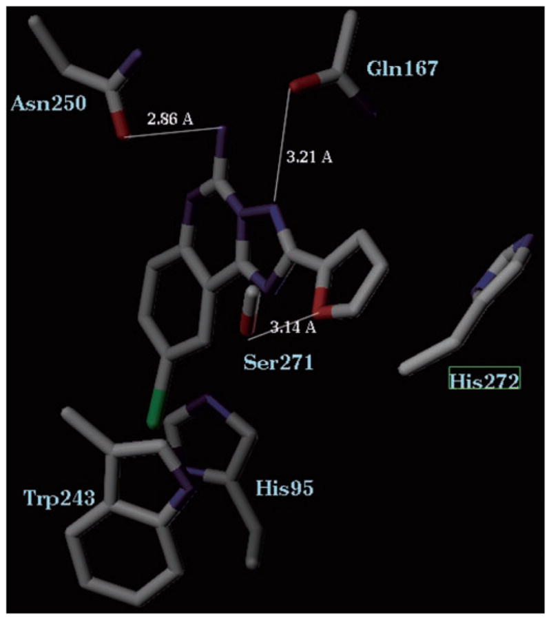
Ligand recognition has been extensively explored in G protein-coupled A(1), A(2A), and A(2B) adenosine receptors but not in the A(3) receptor, which is cerebroprotective and cardioprotective. We mutated several residues of the human A(3) adenosine receptor within transmembrane domains 3 and 6 and the second extracellular loop, which have been predicted by previous molecular modeling to be involved in the ligand recognition, including His(95), Trp(243), Leu(244), Ser(247), Asn(250), and Lys(152). The N250A mutant receptor lost the ability to bind both radiolabeled agonist and antagonist. The H95A mutation significantly reduced affinity of both agonists and antagonists. In contrast, the K152A (EL2), W243A (6.48), and W243F (6.48) mutations did not significantly affect the agonist binding but decreased antagonist affinity by approximately 3-38-fold, suggesting that these residues were critical for the high affinity of A(3) adenosine receptor antagonists. Activation of phospholipase C by wild type (WT) and mutant receptors was measured. The A(3) agonist 2-chloro-N(6)-(3-iodobenzyl)-5'-N-methylcarbamoyladenosine stimulated phosphoinositide turnover in the WT but failed to evoke a response in cells expressing W243A and W243F mutant receptors, in which agonist binding was less sensitive to guanosine 5'-gamma-thiotriphosphate than in WT. Thus, although not important for agonist binding, Trp(243) was critical for receptor activation. The results were interpreted using a rhodopsin-based model of ligand-A(3) receptor interactions.


Figures

Fig. 1
Fig. 1. Heptahelical diagram of the human A3 adenosine receptor
The putative transmembrane domains were modified according to the high resolution rhodopsin model (15). Amino acids mutated in the present study are circled. Residues 286–295 correspond to an extra helical domain in rhodopsin, which is discontinuous from TM7.
Fig. 2
Fig. 2. Sequence alignments of selected regions of A3 adenosine receptors and other G protein-coupled receptors
Residues mutated in this study are shown in boldface type. The standardized numbering system of van Rhee and Jacobson (25) was used to identify residues in the transmembrane domains (TMs) of various receptors. Each residue is identified by two numbers; the first corresponds to the TM in which it is located; the second indicates its position relative to the most conserved residue in that helix, arbitrarily assigned to 50. For example, H3.37 is the histidine in TM3, located 13 residues before the most conserved arginine R3.50; W6.48 corresponds to Trp243.
Fig. 3
Fig. 3
Chemical structures of ligands tested in this study.
Fig. 4
Fig. 4. Effects of mutations in TM3, TM6, and EL2 on the binding of A3 agonist Cl-IB-MECA
The agonist radioligand [125I]I-AB-MECA (1.0 nM) was used in the experiment. The data shown are from a representative example out of at least three independent experiments performed in duplicate.
Fig. 5
Fig. 5. Effects of the Trp mutation on the binding of A3 antagonist MRS1220
The agonist radioligand [125I]I-AB-MECA (1.0 nM) was used in the experiment. The data shown are from a representative example of at least three independent experiments performed in duplicate.
Fig. 6
Fig. 6. Cl-IB-MECA induced phosphoinositide turnover in COS-7 cells expressing WT and mutant human A3 adenosine receptors
Receptors were transiently expressed in COS-7 cells and used 48 h after transfection. The data shown are from a representative example of three independent experiments.
Fig. 7
Fig. 7. Effects of GTP analogue on A3 receptor binding
Shown are the effects of increasing concentrations of GTPγS on binding of the agonist radioligand [125I]I-AB-MECA (1.0 nM) to membranes from COS-7 cells expressing WT and mutant receptors. Results are expressed as percent of binding determined in the absence of GTPγS and are shown as means of values obtained from three experiments performed in duplicate.
Fig. 8
Fig. 8. Molecular model of the nonselective antagonist CGS15943 binding to the human A3 adenosine receptor
Residues in proximity to this triazoloquinazoline are shown. Blue, nitrogen, red, oxygen, green, chlorine.

References

    1. Zhou QY, Li C, Olah ME, Johnson RA, Stiles GL, Civelli O. Proc Natl Acad Sci U S A. 1992;89:7432–7436. - PMC - PubMed
    1. Salvatore SA, Tilley SL, Latour AM, Fletcher DS, Koller BH, Jacobson MA. J Biol Chem. 2000;275:4429–4434. - PubMed
    1. Olah ME, Stiles GL. Pharmacol Ther. 2000;85:55–75. - PubMed
    1. Fredholm BB, Arslan G, Halldner L, Kull B, Schulte G, Wasserman W. Naunyn-Schmiedeberg’s Arch Pharmacol. 2000;362:364–374. - PubMed
    1. Linden J. Annu Rev Pharmacol Toxicol. 2001;41:775–787. - PubMed

Publication types

LinkOut - more resources