. 2002 Mar;128(3):1022-30.
doi: 10.1104/pp.010858.

Ferrous ion transport across chloroplast inner envelope membranes

Affiliations

Ferrous ion transport across chloroplast inner envelope membranes

Richard Shingles et al. Plant Physiol. 2002 Mar.

Abstract

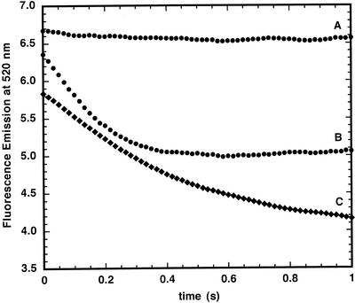
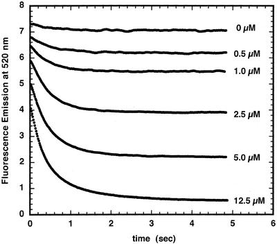
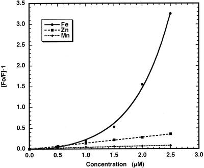
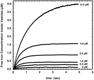
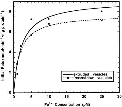
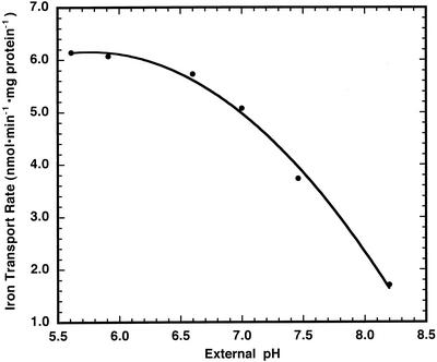
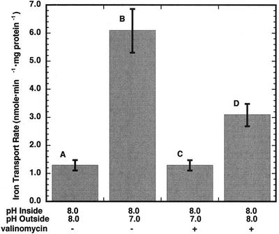
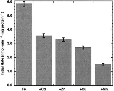
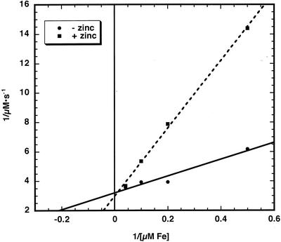
The initial rate of Fe(2+) movement across the inner envelope membrane of pea (Pisum sativum) chloroplasts was directly measured by stopped-flow spectrofluorometry using membrane vesicles loaded with the Fe(2+)-sensitive fluorophore, Phen Green SK. The rate of Fe(2+) transport was rapid, coming to equilibrium within 3s. The maximal rate and concentration dependence of Fe(2+) transport in predominantly right-side-out vesicles were nearly equivalent to those measured in largely inside-out vesicles. Fe(2+) transport was stimulated by an inwardly directed electrochemical proton gradient across right-side-out vesicles, an effect that was diminished by the addition of valinomycin in the presence of K(+). Fe(2+) transport was inhibited by Zn(2+), in a competitive manner, as well as by Cu(2+) and Mn(2+). These results indicate that inward-directed Fe(2+) transport across the chloroplast inner envelope occurs by a potential-stimulated uniport mechanism.


Figures

Figure 1
Figure 1
Ferrous iron quenching of PGSK fluorescence in asolectin and chloroplast inner envelope membrane vesicles. Asolectin and chloroplast inner envelope membrane vesicles were loaded with PGSK using the extrusion method described in “Materials and Methods” (pH = 8.0). Vesicles were rapidly mixed with 5 μm Fe2+ in external buffer (pH 7.0) in a stopped-flow apparatus and fluorescence emission was monitored at 520 nm with excitation at 506 nm. A, Asolectin vesicles. B, Asolectin vesicles pre-incubated 10 min with 20 mm pyrithione. C, Chloroplast inner envelope membrane vesicles.
Figure 2
Figure 2
Ferrous iron quenching of PGSK fluorescence in chloroplast inner envelope membrane vesicles. Chloroplast inner envelope membrane vesicles were loaded with PGSK using the extrusion method described in “Materials and Methods” (pH = 8.0). Vesicles were mixed with varying amounts of Fe2+ in external buffer (pH 7.0) as indicated. Fluorescence emission was monitored at 520 nm after excitation at 506 nm.
Figure 3
Figure 3
Stern-Volmer plot of PGSK fluorescence quenching by cations. The fluorescence emission, monitored at 520 nm with excitation at 506 nm of a solution of 50 μm PGSK in buffer A, was measured in the presence of varying concentrations of Fe2+, Zn2+, or Mn2+.
Figure 4
Figure 4
Intravesicular concentration of ferrous iron in chloroplast inner envelope membrane vesicles loaded with PGSK. Data from Figure 2 were used to determine the intravesicular Fe2+ concentration using the calibration procedure outlined in “Materials in Methods.”
Figure 5
Figure 5
Initial rate of Fe2+ transport in extruded and freeze-/thaw-prepared chloroplast inner envelope membrane vesicles. Inner envelope membranes were prepared by extrusion to produce vesicles largely right side out in orientation. Inner envelope membranes were also prepared by a freeze/thaw technique to produce membranes of largely inside-out orientation. Internal vesicle pH was 8.0, whereas external pH was 7.0. Intravesicular Fe2+ concentration was determined as described in “Materials and Methods” at different concentrations of added Fe2+. Initial rates were determined from the equation describing a single exponential increase.
Figure 6
Figure 6
Effect of external pH on the initial rate of Fe2+ transport across chloroplast inner envelope membrane vesicles. PGSK-loaded inner envelope vesicles (pH = 8.0 inside) were mixed with external buffer at different pH values plus 5 μm Fe2+. PGSK fluorescence quenching was determined over 3 s after mixing and, using the calibration procedure described in “Materials and Methods,” the initial rates of transport were determined.
Figure 7
Figure 7
pH and potential gradient effects on Fe2+ transport across chloroplast inner envelope membrane vesicles. A, Extruded inner envelope vesicles at pH 8.0 were mixed with external buffer at pH 8.0 containing 5 μm Fe2+. B, Extruded inner envelope vesicles at pH 8.0 were mixed with external buffer at pH 7.0 containing 5 μm Fe2+. C, Conditions were the same as B with vesicles pre-incubated with 2 nm valinomycin on ice for 30 min. D, Membrane potential was imposed across the vesicle membranes by mixing vesicles prepared in 100 mm KCl, 10 mm K-HEPES, and 100 mm Suc with an equal volume of the same buffer in which the 100 mm KCl was replaced with 100 mm choline chloride. The instantaneous equilibration of K+ in the presence of valinomycin resulted in a negatively charged vesicle interior. Data shown are the mean ± sd (n = 9).
Figure 8
Figure 8
Effect of added cations on Fe2+ transport across chloroplast inner envelope membrane vesicles. Vesicles contained buffer at pH 8.0 inside and were mixed with external buffer at pH 7.0. Control rate of Fe2+ transport into PGSK-loaded vesicles was established using 5 μm Fe2+. Addition of all other cations was also at 5 μm. Intravesicular Fe2+ concentration was determined as described in “Materials and Methods” at different concentrations of added Fe2+. Initial rates were determined from the equation describing a single exponential increase. Data shown are the mean ±sd (n = 3).
Figure 9
Figure 9
Lineweaver-Burk plot of Zn2+ inhibition of Fe2+ transport. Inner envelope membranes were prepared by extrusion. Intravesicular Fe2+ concentration was determined as described in “Materials and Methods” at different concentrations of Fe2+ in the absence (●) and presence (▪) of 5 μm Zn2+.

References

    1. Bensadoun A, Weinstein D. Assay of proteins in the presence of interfering materials. Anal Biochem. 1976;70:241–250. - PubMed
    1. Berkowitz GA, Peters JS. Chloroplast inner-envelope ATPase acts as a primary H+ pump. Plant Physiol. 1993;102:261–267. - PMC - PubMed
    1. Brüggeman W, Maas-Kantel K, Moog P. Iron uptake by leaf mesophyll cells: the role of the plasma membrane-bound ferric-chelate reductase. Planta. 1993;190:151–155.
    1. Bughio N, Takahashi M, Yoshimura E, Nishizawa NK, Mori S. Light-dependent iron transport into isolated barley chloroplasts. Plant Cell Physiol. 1997;38:101–105.
    1. Cohen CK, Fox TC, Garvin DF, Kochian LV. The role of iron-deficiency stress responses in stimulating heavy-metal transport in plants. Plant Physiol. 1998;116:1063–1072. - PMC - PubMed

Publication types

MeSH terms

LinkOut - more resources