. 2002 Mar 19;99(6):3920-5.
doi: 10.1073/pnas.002024599. Epub 2002 Mar 12.

Complex pattern of Mycobacterium marinum gene expression during long-term granulomatous infection

Affiliations

Complex pattern of Mycobacterium marinum gene expression during long-term granulomatous infection

Kaman Chan et al. Proc Natl Acad Sci U S A. .

Abstract

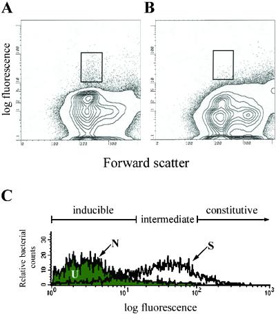
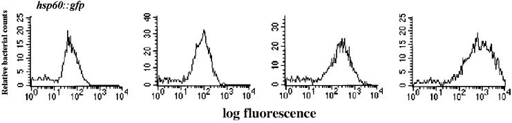
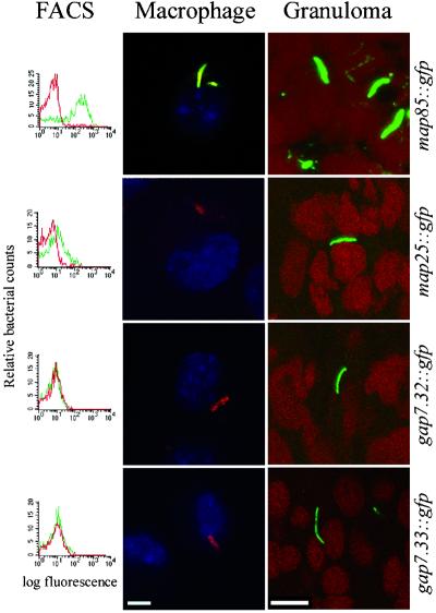
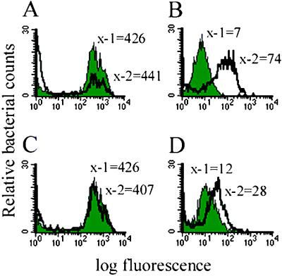
During latent infection of humans with Mycobacterium tuberculosis, bacteria persist in the asymptomatic host within granulomas, organized collections of differentiated macrophages, and other immune cells. The mechanisms for persistence remain poorly understood, as is the metabolic and replicative state of the microbes within granulomas. We analyzed the gene expression profile of Mycobacterium marinum, the cause of fish and amphibian tuberculosis, during its persistence in granulomas. We identified genes expressed specifically when M. marinum persists within granulomas. These granuloma-activated genes were not activated in vitro in response to various conditions postulated to be operant in tuberculous granulomas, suggesting that their granuloma-specific activation was caused by complex conditions that could not be mimicked in vitro. In addition to the granuloma-activated genes, the bacteria resident in granulomas expressed a wide range of metabolic and synthetic genes that are expressed during logarithmic growth in laboratory medium. Our results suggest a dynamic host-pathogen interaction in the granuloma, where metabolically active bacteria are kept in check by the host immune system and where the products of granuloma-specific bacterial genes may thwart the host's attempt to completely eradicate the bacteria.


Figures

Figure 1
Figure 1
Fluorescence profiles of the promoter trap library in infected frogs. (A) Fluorescence profile of bacterial promoters isolated from the spleen tissue of frogs infected with the promoter trap library. (B) Fluorescence profile from the spleen tissue of uninfected frogs. (C) Histograms of the promoter trap library before it had been used to infect frogs (N), from infected frog tissue before sorting for fluorescent bacteria (U), and from infected frog tissue immediately after sorting for fluorescent events (S).
Figure 2
Figure 2
Fluorescence profiles of caps expressed in the granulomas of infected frogs. Histograms of bacterial green fluorescent protein (GFP)-promoter fusions isolated from the tissue of infected frogs that retain fluorescence equal to or greater than an hsp60∷GFP fusion when plated on bacterial media. Represented are fluorescence profiles of three caps with different fluorescence intensities.
Figure 3
Figure 3
Analysis of individual promoter fusions. Individual promoter fusions isolated from frog granulomas in the library screen were analyzed for activity in DMEM, J774 macrophages, and granulomas. Red lines in the FACS histograms represent fluorescence in DMEM; green lines indicate fluorescence in macrophages. Macrophage confocal images are overlays of red (bacterial membrane protein), green [for expression of the green fluorescent protein (GFP) fusion in the promoter constructs], and blue (macrophage nuclear staining). Therefore GFP expression in macrophages renders the bacteria yellow. In the absence of GFP expression, they are red fluorescent. Granuloma confocal images are overlays of red (macrophage nuclear stain) and green (for expression of the GFP promoter fusion). The maps and gaps have minimal fluorescence in laboratory medium. map 85 is fluorescent in both macrophages (yellow) and granulomas (green), whereas the two gaps are fluorescent only in granulomas (green) and not in macrophages (red). map 25 straddles the classification between a map and a gap, being only slightly activated in macrophages (red with faint yellow) and green in the granuloma. (Bars represent 10 microns.)
Figure 4
Figure 4
Fluorescence profiles of promoters under in vitro inducing conditions. Shaded peaks correspond to control conditions (pH 7.0 and 200 μM Mg2+) whereas unshaded peaks correspond to inducing conditions (either pH 6.25 or 20 μM Mg2+). x-1 and x-2 represent the median fluorescence of bacteria in each of the two conditions. (A and B) The msp12 constitutive promoter and the map 24 promoter at pH 7.0 and pH 6.25, respectively. (C and D) The msp12 constitutive promoter and map 25 at 200 μM and 20 μM Mg2+, respectively.

References

    1. Dannenberg A M., Jr Hospital Practice. 1993;28:51–58. - PubMed
    1. Flynn J L, Chan J. Infect Immun. 2001;69:4195–4201. - PMC - PubMed
    1. Manabe Y C, Bishai W R. Nat Med. 2000;6:1327–1329. - PubMed
    1. Wayne L G, Sohaskey C D. Annu Rev Microbiol. 2001;55:139–163. - PubMed
    1. Glickman M S, Jacobs W R., Jr Cell. 2001;104:477–485. - PubMed

Publication types

MeSH terms