. 2002 Mar 19;99(6):3615-20.
doi: 10.1073/pnas.062030799. Epub 2002 Mar 12.

Restoration of fragile histidine triad (FHIT) expression induces apoptosis and suppresses tumorigenicity in lung and cervical cancer cell lines

Affiliations

Restoration of fragile histidine triad (FHIT) expression induces apoptosis and suppresses tumorigenicity in lung and cervical cancer cell lines

Luca Roz et al. Proc Natl Acad Sci U S A. .

Abstract

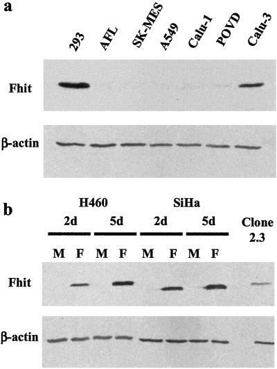
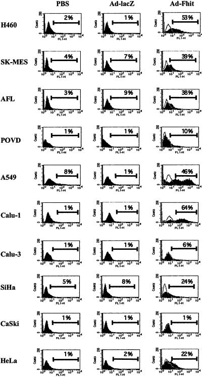
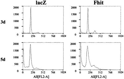
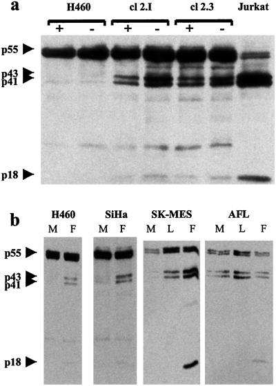
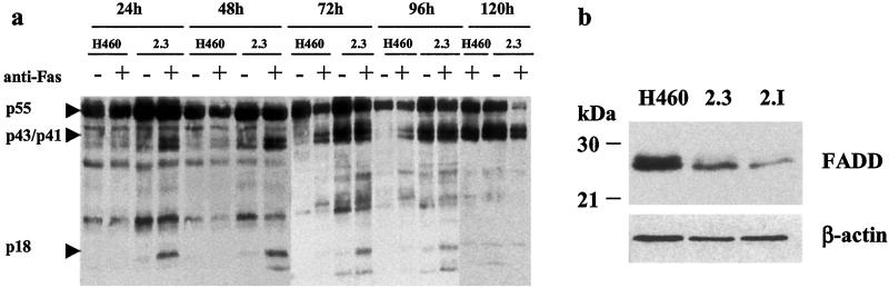
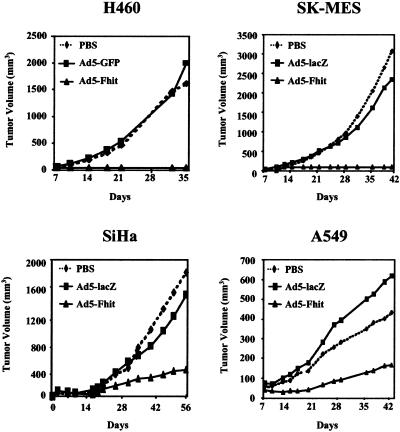
Loss of expression of the Fhit protein is often associated with the development of many human epithelial cancers, including lung and cervical carcinomas. Restoration of Fhit expression in cell lines derived from these tumors has however yielded conflicting results, prompting the need for careful evaluation of the oncosuppressive potential of FHIT. In the present study, we have investigated the effect of Fhit reintroduction in seven lung cancer and three cervical cancer cell lines. To achieve efficient gene transfer and high levels of transgene expression, we have used an adenoviral vector to transduce the FHIT gene. The induction of apoptosis was evaluated by using the terminal deoxynucleotidyltransferase-mediated dUTP nick end labeling assay and propidium iodide staining. Activation of caspases was detected by using Western blot analysis, and tumorigenic potential of transduced cells in the nude mouse was also assessed. Restoration of Fhit expression induced apoptosis in all Fhit-negative cell lines, with Calu-1, H460, and A549 being the most susceptible among the lung cancer cell lines and SiHa cells among cervical carcinomas. Activation of caspase-8 was always associated with Fhit-mediated apoptosis, and in vivo tumorigenicity was either abolished by FHIT gene transfer (in H460 and SK-Mes cells) or strongly suppressed (in A549 and SiHa cells). Our data demonstrate oncosuppressive properties and strong proapoptotic activity of the Fhit protein in lung and cervical cancer cell lines and strengthens the hypothesis of its possible use as a therapeutic tool.


Figures

Figure 1
Figure 1
Endogenous Fhit expression in cancer cell lines and effect of adenoviral Fhit transfer. (a) Immunoblot analysis of Fhit expression in lung cancer cell lines. 293, Human embryonic kidney cells used as positive control. (b) Fhit expression in cell lines derived from lung (H460) and cervical (SiHa) carcinomas 2 (2d) and 5 (5d) days after treatment with Ad5-Fhit (lanes F) compared with endogenous Fhit levels in mock infected cells (lanes M). 2.3, Stable FHIT-transfectant of H460 cells used as positive control for high levels of Fhit expression.
Figure 2
Figure 2
Flow cytometry evaluation of TUNEL analysis of Fhit-induced apoptosis. For each cell line, the fluorescence profile of mock infected cells (PBS) or cells treated with control adenovirus (Ad-lacZ) or with Fhit-expressing adenoviral vector (Ad-Fhit) is presented. Percentage of apoptotic cells (cells displaying increased fluorescence compared with controls) is indicated above the marker bar in each panel. In the profile of Ad5-Fhit-treated cells, the negative control represented by cells incubated in the absence of terminal transferase enzyme is also shown (open graph). Results shown are representative examples of experiments repeated at least twice, in which the SD was <15%.
Figure 3
Figure 3
Effects of Fhit reexpression on the cell cycle. Distribution of H460 cells in the cell cycle was analyzed by propidium iodide staining. The percentage of cells in G0/G1, S, and G2 was, respectively, 81.6%, 12.4%, and 6% for Ad5-lacZ-infected cells (lacZ) and 80.2%, 6.6%, and 13.2% for Ad5-Fhit-infected cells (Fhit) 3 days postinfection (3d) and 84.4%, 10.1%, and 5.5% (lacZ) and 71.5%, 27.5%, and 1% (Fhit) 5 days postinfection (5d).
Figure 4
Figure 4
Restored Fhit expression induces caspase-8 activation. (a) H460 cells and its stable Fhit transfectants clone 2.I and 2.3 were kept in normal culture conditions (10% FCS, +) or serum starved (−) and then analyzed by Western blot by using a caspase-8-specific monoclonal antibody. Presence of cleavage forms of pro-caspase (p43/p41 and the active p18 subunit) indicates activation. (b) Indicated cell lines were infected with adenoviral vectors expressing Fhit (F), lacZ (L), or mock-infected (M) and analyzed after 5 days.
Figure 5
Figure 5
(a) Fhit-expression modulates apoptotic response to Fas treatment. H460 cell line and its stable Fhit-transfectant (clone 2.3) were cultured in the presence (+) or absence (−) of an agonistic anti-Fas antibody (CH-11). At the indicated time points, cells were harvested and processed for immunoblotting with the anti-caspase-8 monoclonal antibody C-15. Cleavage bands (p43/41 and p18), indicating caspase-8 activation, are already detected in Fhit-expressing cells after 24 h, and, after 120 h, the inactive form of caspase-8 is almost entirely converted into the active subunits. Toxic effects of the CH-11 antibody are also visible in H460 cells after prolonged treatment, confirming that the Fas pathway is functional in these cells and suggesting that the higher sensitivity of the clones to Fas-induced apoptosis could be due to a direct effect of Fhit on the apoptotic signal transduction or on the apoptotic threshold. (b) Down-regulation of FADD in stable Fhit-transfectants. Western blot showing reduced expression of FADD in stable Fhit transfectants (clones 2.3 and 2.I) compared with H460 parental cell line. β-actin, same blot hybridized with anti-actin antibody.
Figure 6
Figure 6
Adenoviral-mediated Fhit expression suppresses in vivo tumorigenicity in lung and cervical cancer cell lines. Cell lines were infected in vitro with Ad5-Fhit, control adenoviruses (Ad5-GFP or Ad5-lacZ), or mock infected (PBS) and then injected s.c. into nude mice (four to six animals per group). All experiments were repeated at least twice, and representative results are reported as the mean for each treatment group. Although variability in tumor sizes was observed in some treatment groups, Fhit expression completely suppressed tumorigenicity of H460 and SK-MES cell lines, and the differences between the tumor volumes in the groups injected with control vector-treated cells and mock-infected cells were not significant (Student's t test, two-tail; P = 0.39 for H460, P = 0.27 for SK-MES, P = 0.37 for SiHa, and P = 0.34 for A549 cells). A significant reduction (P < 0.05) of tumor size was observed in Ad5-Fhit-treated SiHa and A549 cells compared with untreated (P = 0.008 for SiHa and P = 0.025 for A549) or Ad5-lacZ-treated cells (P = 0.001 for SiHa and P = 0.022 for A549 cells).

References

    1. Ohta M, Inoue H, Cotticelli M G, Kastury K, Baffa R, Palazzo J, Siprashvili Z, Mori M, McCue P, Druck T, et al. Cell. 1996;84:587–597. - PubMed
    1. Sozzi G, Veronese M L, Negrini M, Baffa R, Cotticelli M G, Inoue H, Tornielli S, Pilotti S, DeGregario L, Pastorino V, Pierotti M A, et al. Cell. 1996;85:17–26. - PubMed
    1. Sozzi G, Pastorino U, Moiraghi L, Tagliabue E, Pezzella F, Ghirelli C, Tornielli S, Sard L, Huebner K, Pierotti M A, et al. Cancer Res. 1998;58:5032–5037. - PubMed
    1. Baffa R, Gomella L G, Vecchione A, Bassi P, Mimori K, Sedor J, Calviello C M, Gardiman M, Minimo C, Strup S E, et al. Am J Pathol. 2000;156:419–424. - PMC - PubMed
    1. Campiglio M, Pekarsky Y, Menard S, Tagliabue E, Pilotti S, Croce C M. Cancer Res. 1999;59:3866–3869. - PubMed

Publication types

MeSH terms