

Deletion of the thyroid hormone receptor alpha 1 prevents the structural alterations of the cerebellum induced by hypothyroidism
- PMID: 11891331
- PMCID: PMC122635
- DOI: 10.1073/pnas.062413299
Deletion of the thyroid hormone receptor alpha 1 prevents the structural alterations of the cerebellum induced by hypothyroidism
Abstract
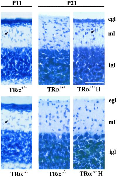
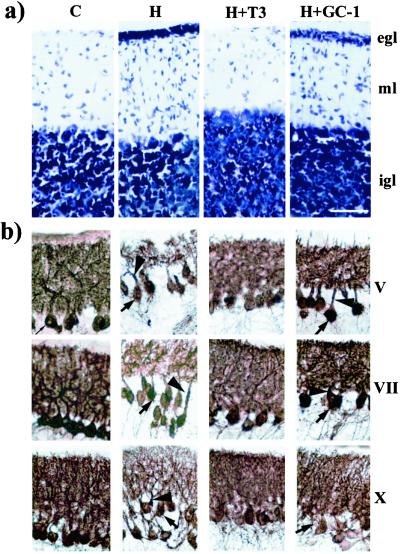
Thyroid hormone (T3) controls critical aspects of cerebellar development, such as migration of postmitotic granule cells and terminal differentiation of Purkinje cells. T3 acts through nuclear receptors (TR) of two types, TRalpha1 and TRbeta, that either repress or activate gene expression. We have analyzed the cerebellar structure of developing mice lacking the TRalpha1 isoform, which normally accounts for about 80% of T3 receptors in the cerebellum. Contrary to what was expected, granule cell migration and Purkinje cell differentiation were normal in the mutant mice. Even more striking was the fact that when neonatal hypothyroidism was induced, no alterations in cerebellar structure were observed in the mutant mice, whereas the wild-type mice showed delayed granule cell migration and arrested Purkinje cell growth. The results support the idea that repression by the TRalpha1 aporeceptor, and not the lack of thyroid hormone, is responsible for the hypothyroid phenotype. This conclusion was supported by experiments with the TRbeta-selective compound GC-1. Treatment of hypothyroid animals with T3, which binds to TRalpha1 and TRbeta, prevents any defect in cerebellar structure. In contrast, treatment with GC-1, which binds to TRbeta but not TRalpha1, partially corrects Purkinje cell differentiation but has no effect on granule cell migration. Our data indicate that thyroid hormone has a permissive effect on cerebellar granule cell migration through derepression by the TRalpha1 isoform.
Figures


Similar articles
-
Thyroid hormone induces cerebellar Purkinje cell dendritic development via the thyroid hormone receptor alpha1.J Neurosci. 2003 Nov 19;23(33):10604-12. doi: 10.1523/JNEUROSCI.23-33-10604.2003. J Neurosci. 2003. PMID: 14627645 Free PMC article.
-
Influence of thyroid hormone and thyroid hormone receptors in the generation of cerebellar gamma-aminobutyric acid-ergic interneurons from precursor cells.Endocrinology. 2007 Dec;148(12):5746-51. doi: 10.1210/en.2007-0567. Epub 2007 Aug 30. Endocrinology. 2007. PMID: 17761765
-
Requirement for thyroid hormone receptor beta in T3 regulation of cholesterol metabolism in mice.Mol Endocrinol. 2002 Aug;16(8):1767-77. doi: 10.1210/me.2002-0009. Mol Endocrinol. 2002. PMID: 12145333
-
Isoform-dependent actions of thyroid hormone nuclear receptors: lessons from knockin mutant mice.Steroids. 2005 May-Jun;70(5-7):450-4. doi: 10.1016/j.steroids.2005.02.003. Epub 2005 Mar 16. Steroids. 2005. PMID: 15862829 Review.
-
Current perspectives on the role of thyroid hormone in growth and development of cerebellum.Cerebellum. 2003;2(4):279-89. doi: 10.1080/14734220310011920. Cerebellum. 2003. PMID: 14964687 Review.
Cited by
-
Redox activation of JNK2α2 mediates thyroid hormone-stimulated proliferation of neonatal murine cardiomyocytes.Sci Rep. 2019 Nov 27;9(1):17731. doi: 10.1038/s41598-019-53705-1. Sci Rep. 2019. PMID: 31776360 Free PMC article.
-
Thyroid hormone action during GABAergic neuron maturation: The quest for mechanisms.Front Endocrinol (Lausanne). 2023 Oct 3;14:1256877. doi: 10.3389/fendo.2023.1256877. eCollection 2023. Front Endocrinol (Lausanne). 2023. PMID: 37854197 Free PMC article. Review.
-
Retarded developmental expression and patterning of retinal cone opsins in hypothyroid mice.Endocrinology. 2009 Mar;150(3):1536-44. doi: 10.1210/en.2008-1092. Epub 2008 Oct 30. Endocrinology. 2009. PMID: 18974269 Free PMC article.
-
Thyroid hormone receptor alpha is a molecular switch of cardiac function between fetal and postnatal life.Proc Natl Acad Sci U S A. 2004 Jul 13;101(28):10332-7. doi: 10.1073/pnas.0401843101. Epub 2004 Jul 6. Proc Natl Acad Sci U S A. 2004. PMID: 15240882 Free PMC article.
-
Polychlorinated biphenyls (Aroclor 1254) do not uniformly produce agonist actions on thyroid hormone responses in the developing rat brain.Endocrinology. 2008 Aug;149(8):4001-8. doi: 10.1210/en.2007-1774. Epub 2008 Apr 17. Endocrinology. 2008. PMID: 18420739 Free PMC article.
References
-
- Izumo S, Mahdavi V. Nature (London) 1988;334:539–542. - PubMed
-
- Koening R J, Lazar M A, Holdin R A, Brent G A, Larsen P R, Chin W W, Moore D D. Nature (London) 1989;337:659–661. - PubMed
-
- Chassande O, Fraichard A, Gauthier K, Flamant F, Legrand C, Savatier P, Laudet V, Samarut J. Mol Endocrinol. 1997;11:1278–1290. - PubMed
Publication types
MeSH terms
Substances
Grants and funding
LinkOut - more resources
Full Text Sources
Medical
Molecular Biology Databases
Miscellaneous
