

Induction of p57(KIP2) expression by p73beta
- PMID: 11891335
- PMCID: PMC122557
- DOI: 10.1073/pnas.062491899
Induction of p57(KIP2) expression by p73beta
Abstract
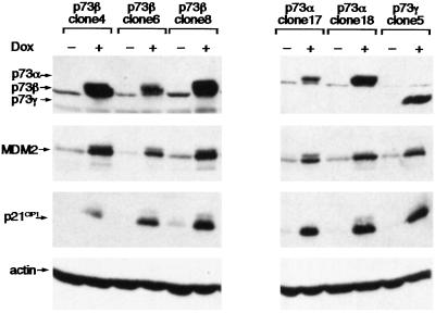
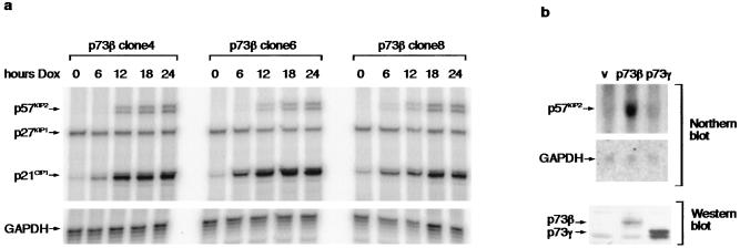
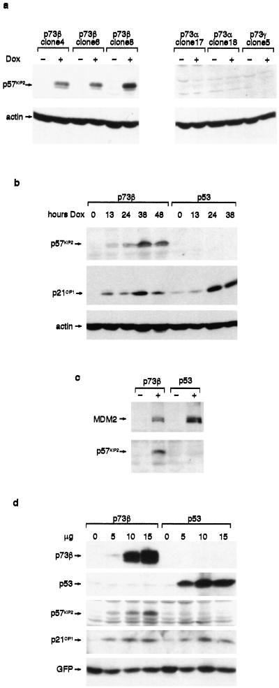
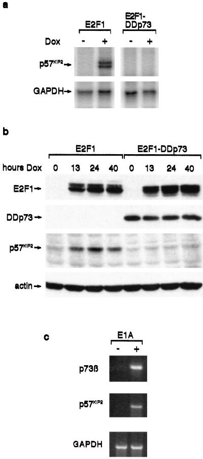
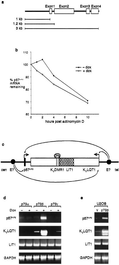
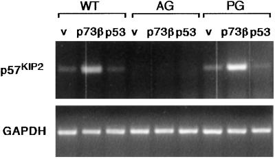
The p53-related protein p73 has many functions similar to that of p53 including the ability to induce cell-cycle arrest and apoptosis. Both p53 and p73 function as transcription factors, and p73 activates expression of many genes that also are regulated by p53. Despite their similarities, it is evident that p53 and p73 are not interchangeable functionally, with p73 playing a role in normal growth and development that is not shared by p53. In this paper we describe the ability of p73beta but not p53 to activate expression of the cyclin-dependent kinase inhibitor p57(KIP) and KvLQT1, two genes that are coregulated in an imprinted region of the genome. Our results suggest that p73 may regulate expression of genes through mechanisms that are not shared by p53, potentially explaining the different contributions of p53 and p73 to normal development.
Figures
Similar articles
-
Multiple mechanisms regulate imprinting of the mouse distal chromosome 7 gene cluster.Mol Cell Biol. 1998 Jun;18(6):3466-74. doi: 10.1128/MCB.18.6.3466. Mol Cell Biol. 1998. PMID: 9584186 Free PMC article.
-
Induction of p73beta by a naphthoquinone analog is mediated by E2F-1 and triggers apoptosis in HeLa cells.FEBS Lett. 2002 Jul 3;522(1-3):161-7. doi: 10.1016/s0014-5793(02)02921-6. FEBS Lett. 2002. PMID: 12095638
-
A role of p73 in mitotic exit.J Biol Chem. 2005 Aug 26;280(34):30354-60. doi: 10.1074/jbc.M500635200. Epub 2005 Jun 28. J Biol Chem. 2005. PMID: 15985436
-
From p63 to p53 across p73.FEBS Lett. 2001 Feb 16;490(3):163-70. doi: 10.1016/s0014-5793(01)02119-6. FEBS Lett. 2001. PMID: 11223031 Review.
-
Regulation and activation of p53 and its family members.Cell Death Differ. 1999 Dec;6(12):1162-8. doi: 10.1038/sj.cdd.4400625. Cell Death Differ. 1999. PMID: 10637431 Review.
Cited by
-
Imprinted Zac1 in neural stem cells.World J Stem Cells. 2015 Mar 26;7(2):300-14. doi: 10.4252/wjsc.v7.i2.300. World J Stem Cells. 2015. PMID: 25815116 Free PMC article. Review.
-
Modification of the ionizing radiation response in living cells by an scFv against the DNA-dependent protein kinase.Nucleic Acids Res. 2003 Oct 15;31(20):5848-57. doi: 10.1093/nar/gkg775. Nucleic Acids Res. 2003. PMID: 14530433 Free PMC article.
-
Hsp72 mediates TAp73α anti-apoptotic effects in small cell lung carcinoma cells.J Cell Mol Med. 2011 Aug;15(8):1757-68. doi: 10.1111/j.1582-4934.2010.01166.x. J Cell Mol Med. 2011. PMID: 20807285 Free PMC article.
-
p53 Family and Cellular Stress Responses in Cancer.Front Oncol. 2014 Oct 21;4:285. doi: 10.3389/fonc.2014.00285. eCollection 2014. Front Oncol. 2014. PMID: 25374842 Free PMC article. Review.
-
Tbx2 controls lung growth by direct repression of the cell cycle inhibitor genes Cdkn1a and Cdkn1b.PLoS Genet. 2013;9(1):e1003189. doi: 10.1371/journal.pgen.1003189. Epub 2013 Jan 17. PLoS Genet. 2013. PMID: 23341776 Free PMC article.
References
-
- Kaelin W G., Jr Oncogene. 1999;18:7701–7705. - PubMed
-
- Yang A, Walker N, Bronson R, Kaghad M, Oosterwegel M, Bonnin J, Vagner C, Bonnet H, Dikkes P, Sharpe A, McKeon F, Caput D. Nature (London) 2000;404:99–103. - PubMed
-
- Yang A, Schweitzer R, Sun D, Kaghad M, Walker N, Bronson R T, Tabin C, Sharpe A, Caput D, Crum C, McKeon F. Nature (London) 1999;398:714–717. - PubMed
-
- Mills A A, Zheng B, Wang X-J, Vogel H, Roop D R, Bradley A. Nature (London) 1999;398:708–713. - PubMed
-
- Kawano S, Miller C W, Gombart A F, Bartram C R, Matsuo Y, Asou H, Sakashita A, Said J, Tatsumi E, Koeffler H P. Blood. 1999;94:1113–1120. - PubMed
MeSH terms
Substances
LinkOut - more resources
Full Text Sources
Other Literature Sources
Molecular Biology Databases
Research Materials
Miscellaneous
