. 2002 Mar 19;99(6):3944-9.
doi: 10.1073/pnas.062652199. Epub 2002 Mar 12.

Nuclear entry and CRM1-dependent nuclear export of the Rous sarcoma virus Gag polyprotein

Affiliations

Nuclear entry and CRM1-dependent nuclear export of the Rous sarcoma virus Gag polyprotein

Lisa Z Scheifele et al. Proc Natl Acad Sci U S A. .

Abstract

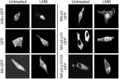
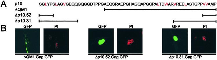
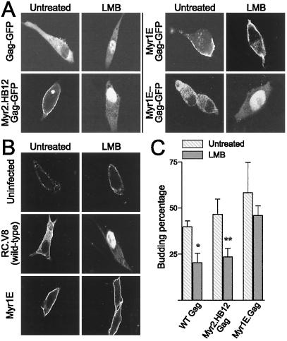
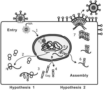
The retroviral Gag polyprotein directs budding from the plasma membrane of infected cells. Until now, it was believed that Gag proteins of type C retroviruses, including the prototypic oncoretrovirus Rous sarcoma virus, were synthesized on cytosolic ribosomes and targeted directly to the plasma membrane. Here we reveal a previously unknown step in the subcellular trafficking of the Gag protein, that of transient nuclear localization. We have identified a targeting signal within the N-terminal matrix domain that facilitates active nuclear import of the Gag polyprotein. We also found that Gag is transported out of the nucleus through the CRM1 nuclear export pathway, based on observations that treatment of virus-expressing cells with leptomycin B resulted in the redistribution of Gag proteins from the cytoplasm to the nucleus. Internal deletion of the C-terminal portion of the Gag p10 region resulted in the nuclear sequestration of Gag and markedly diminished budding, suggesting that the nuclear export signal might reside within p10. Finally, we observed that a previously described matrix mutant, Myr1E, was insensitive to the effects of leptomycin B, apparently bypassing the nuclear compartment during virus assembly. Myr1E has a defect in genomic RNA packaging, implying that nuclear localization of Gag might be involved in viral RNA interactions. Taken together, these findings provide evidence that nuclear entry and egress of the Gag polyprotein are intrinsic components of the Rous sarcoma virus assembly pathway.


Figures

Figure 1
Figure 1
The RSV Gag protein and derivatives. The RSV Gag protein (Pr76) is depicted at the top, with cleavage proteins MA, p2a, p2b, p10, CA, NC, and PR indicated. RC.Myr0 is a derivative of pRCV8 and expresses the wild-type Prague C MA protein (45). Gag-GFP fusion proteins and truncations of MA fused to GFP are depicted below. Note that GFP replaces the PR sequence in Gag-GFP (ΔPR). Gag-GFP substitution mutants are shown in the bottom panel. The box labeled “Src” contains the N-terminal 10 residues of the Src oncoprotein. Myristic acid is shown as a zigzag line.
Figure 2
Figure 2
Subcellular localization of the RSV MA protein. (A Upper) Subcellular localizations of GFP, MA-GFP, and Gag-GFP fusion proteins were analyzed in live cells using confocal microscopy 18 h after transfection. (Lower) Transfected QT6 cells were fixed and incubated with polyclonal anti-MA Ab and Cy3-conjugated secondary Ab. (B) Immunoblot analysis of intracellular expression levels of indicated proteins from lysates of transfected cells by using an anti-GFP Ab. Molecular weight markers are indicated to the left. (C) Mapping a nuclear-targeting sequence within the MA domain. Plasmids encoding truncations of the MA sequence fused to GFP were transfected and cells fixed, and epifluorescence was detected. The cells were stained with propidium iodide to visualize nuclear DNA, and identical fields are shown.
Figure 3
Figure 3
Identification of an LMB-sensitive NES within the Gag protein. Live cells transfected with plasmids encoding C-terminal truncations of Gag fused to GFP were examined by confocal microscopy. LMB-treated cells were incubated with 10 ng/ml LMB for 2 h before imaging.
Figure 4
Figure 4
Effects of p10 deletions on subcellular localization. (A) Schematic diagram of the p10 sequence with hydrophobic residues that could function as an NES highlighted in red. Gag mutants with p10 deletions are shown below with heavy black lines indicating residues present in the mutant protein. (B) Confocal micrographs of cells expressing Gag-GFP p10 mutants reveal a putative NES. At 18 h after transfection, cells were fixed in paraformaldehyde. (Left) GFP epifluorescence. (Right) Propidium iodide-stained nuclei.
Figure 5
Figure 5
LMB sensitivity of mutant Gag proteins with defects in RNA packaging and dimerization. (A) Live cells expressing the indicated Gag-GFP proteins were visualized without (Left) and with (Right) LMB treatment, revealing that wild-type Gag and mutants Myr2.HB12 and Myr1E− become trapped in the nucleus after treatment whereas Myr1E is insensitive to the effects of the drug. (B) Gag localization was analyzed in cells stably expressing wild-type or mutant RSV proviral genomes by indirect immunofluorescence in fixed cells either untreated or treated with LMB. Note that patterns of subcellular localization were the same as in A, indicating that GFP had not influenced the distribution of Gag. (C) Particle release in response to LMB treatment. The amount of immunoprecipitated Gag protein released from transfected cells during a 2.5-h-labeling period was divided by the total Gag protein detected in cells and medium to calculate budding percentage. Budding percentages from untreated (hatched bars) and treated (solid gray bars) cells were compared. Treated cells were incubated with LMB for 1–2 h followed by metabolic radiolabeling for 2.5 h in the presence of LMB. Each bar represents the average of three independent experiments with SDs indicated. Inhibition of particle assembly with LMB treatment was determined by a two-sample t test; *, P = 0.0049 and **, P = 0.014.
Figure 6
Figure 6
Model for the role of the nuclear localization of Gag during RSV replication. Hypothesis 1 (steps 1–4): The nuclear-targeting signal within MA delivers the cleaved MA protein to the nucleus during viral entry. Nuclear Gag proteins are exported via the CRM1-dependent NES so they are available in the cytoplasm to direct particle assembly. Hypothesis 2: (steps 5–7) The nuclear-targeting signal carries the Gag protein into the nucleus where Gag might interact with unspliced viral RNA to begin the packaging process. Cytoplasmic relocalization of Gag occurs via the NES; Gag–RNA complexes provide a nucleation point for Gag multimerization, and assembly intermediates are transported to the plasma membrane where budding occurs. Notably, hypotheses 1 and 2 are not mutually exclusive.

Similar articles

Cited by

References

    1. Whittaker G R, Helenius A. Virology. 1998;246:1–23. - PubMed
    1. Hatzoiioannou T G, Goff S P. J Virol. 2001;75:9526–9531. - PMC - PubMed
    1. Cullen B R. Mol Cell Biol. 2000;20:4181–4187. - PMC - PubMed
    1. Ogert R A, Beemon K L. J Virol. 1998;72:3407–3411. - PMC - PubMed
    1. Mattaj I W, Englmeier L. Annu Rev Biochem. 1998;67:265–306. - PubMed

Publication types

MeSH terms