. 2002 Jan;7(1):6-16.
doi: 10.1379/1466-1268(2002)007<0006:xshsph>2.0.co;2.

Xenopus small heat shock proteins, Hsp30C and Hsp30D, maintain heat- and chemically denatured luciferase in a folding-competent state

Affiliations

Xenopus small heat shock proteins, Hsp30C and Hsp30D, maintain heat- and chemically denatured luciferase in a folding-competent state

Rashid Abdulle et al. Cell Stress Chaperones. 2002 Jan.

Abstract

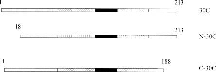
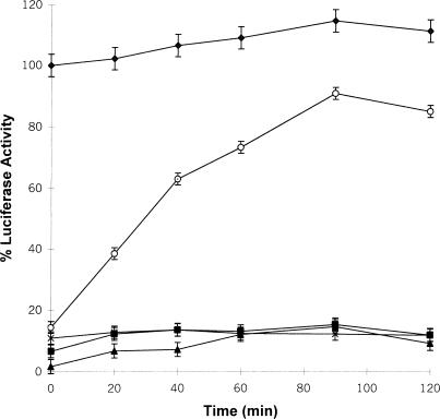
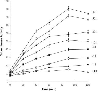
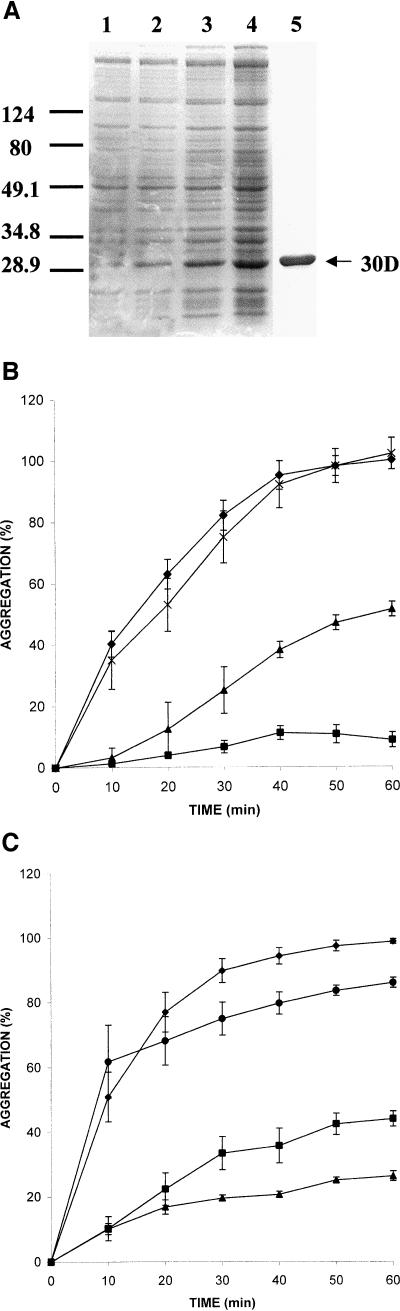
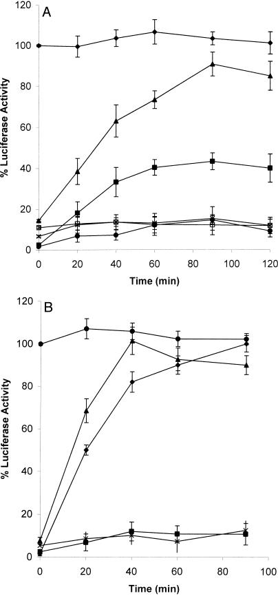
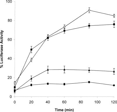
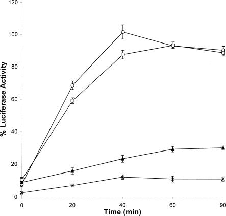
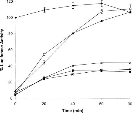
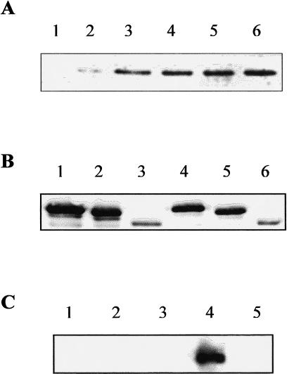
In this study we characterized the chaperone functions of Xenopus recombinant Hsp30C and Hsp30D by using an in vitro rabbit reticulocyte lysate (RRL) refolding assay system as well as a novel in vivo Xenopus oocyte microinjection assay. Whereas heat- or chemically denaturated luciferase (LUC) did not regain significant enzyme activity when added to RRL or microinjected into Xenopus oocytes, compared with native LUC, denaturation of LUC in the presence of Hsp30C resulted in a reactivation of enzyme activity up to 80-100%. Recombinant Hsp30D, which differs from Hsp30C by 19 amino acids, was not as effective as its isoform in preventing LUC aggregation or maintaining it in a folding-competent state. Removal of the first 17 amino acids from the N-terminal region of Hsp30C had little effect on its ability to maintain LUC in a folding-competent state. However, deletion of the last 25 residues from the C-terminal end dramatically reduced Hsp30C chaperone activity. Coimmunoprecipitation and immunoblot analyses revealed that Hsp30C remained associated with heat-denatured LUC during incubation in reticulocyte lysate and that the C-terminal mutant exhibited reduced affinity for unfolded LUC. Finally, we found that Hsc70 present in RRL interacted only with heat-denatured LUC bound to Hsp30C. These findings demonstrate that Xenopus Hsp30 can maintain denatured target protein in a folding-competent state and that the C-terminal end is involved in this function.


Figures

Fig 1.
Fig 1.
A diagram of Xenopus Hsp30C and mutants. The N-terminal mutant N-30C lacking the first 17 amino acids and the C-terminal mutant C-30C with the last 25 amino acids deleted were created by polymerase chain reaction–directed mutagenesis. The textured region is the estimated α-crystallin domain, whereas the α-crystallin consensus sequence is contained within the black rectangle
Fig 2.
Fig 2.
Luciferase (LUC) heat denatured in the presence of recombinant Hsp30C (30C) can be reactivated in vitro by reticulocyte lysate (RRL). LUC (0.2 μM) was incubated with 6.0 μM of 30C (○) or bovine serum albumin (x), and heat denatured at 42°C or kept at 22°C (♦) for 15 minutes. Samples were adjusted to 60 % RRL and 6 μM adenosine triphosphate (ATP), and incubated at 30°C for various periods of time. LUC activity was assayed as described in “Materials and Methods.” Some samples were heat treated without 30C (▪) or assayed without ATP (▴). The data are calculated as a percentage of the activity of unheated LUC at 0 minute, and expressed as the mean of 4–6 trials ± SE
Fig 3.
Fig 3.
The effect of different amounts of recombinant Hsp30C (30C) during heat denaturation on luciferase (LUC) enzyme reactivation in vitro. LUC (0.2 μM) was heat denatured at 42°C with various molar quantities at 30C-LUC molar ratios, as indicated beside each curve. LUC activity was assayed after incubation with reticulocyte lysate and adenosine triphosphate, as described in “Materials and Methods.” All data are expressed as a percentage of the activity of native LUC and shown as the mean ± SE
Fig 4.
Fig 4.
Luciferase (LUC) heat denatured in the presence of recombinant Hsp30C (30C) can be refolded in vivo after microinjection into Xenopus oocytes. LUC (0.2 μM) was incubated at 22°C (♦), or heat denatured alone at 42°C (X) or in the presence of either 6 μM bovine serum albumin (•) or 30C at 30C-LUC molar ratios of 1:1 (□), 10:1 (▴), or 30:1 (▵) for 15 minutes. Mixtures (containing 1.38 fmol of LUC in 26.7 nL) were microinjected into Xenopus oocytes, and LUC activity in the oocytes was monitored over time as described in “Materials and Methods.” Data are representative of 3–5 trials and shown as the mean ± SE
Fig 5.
Fig 5.
Molecular chaperone activity of Xenopus Hsp30D. (A) Expression and purification of Hsp30D recombinant protein. Total bacterial protein from E coli BL21(DE3) cells containing the Hsp30D-pRSETB expression vector was collected before (lane 1) and after 5 hours, 14 hours, and 20 hours (lanes 2–4) of isopropyl-β-d-thiogalactopyranoside treatment. Recombinant protein was purified by means of nickel affinity column chromatography, as detailed in Fernando and Heikkila (2000). Protein was analyzed by sodium dodecyl sulfate–polyacrylamide gel electrophoresis and visualized by Coomassie Brilliant Blue staining. Five micrograms of purified recombinant Hsp30D (30D) is shown in lane 5. Molecular mass markers in kDa are indicated on the left side of the figure. (B) Prevention of heat-induced citrate synthase (CS) aggregation by 30D. Aggregation assays were carried out using the protocols described previously (Fernando and Heikkila 2000). CS or luciferase (LUC) at 150 nM monomer concentrations was mixed with various molar amounts of 30D protein or incubated alone in a 50 mM N-2-hydroxythylpiperazine-N′-2-ethane-sulfonic acid–KOH (pH 7.5) buffer and heated at 42°C. Light scattering was determined at 10-minute intervals in a Beckman DU7 spectrophotometer at 320 nm. An increase in absorbance was indicative of protein aggregation. Data are representative of 4–6 trials and were calculated as a percentage of the maximum aggregation of CS or LUC after 60 minutes and expressed as the mean ± SE. CS was heat treated alone (♦, 0.1 μM) or in the presence of either 30D (▴, 0.1 μM; ▪, 0.5 μM) or IgG (x, 0.5 μM). (C) Inhibition of heat-induced aggregation of LUC by 30D. LUC was heat treated alone (♦, 0.1 μM) or in the presence of 30D (▪, 0.5 μM; ▴, 1.0 μM) or IgG (•, 0.5 μM)
Fig 6.
Fig 6.
In vitro and in vivo enzyme reactivation of luciferase (LUC) heat denatured in the presence of Hsp30D. LUC refolding assays were carried out as described in the legends of Figures 2 and 4. In the reticulocyte lysate (RRL) refolding assays (panel A), LUC (0.2 μM) was maintained at 22°C (♦) or combined with 6.0 μM of recombinant Hsp30D (30D) (▪), 30C (▴), or bovine serum albumin (BSA) (□), and heat denatured at 42°C for 15 minutes prior to incubation with RRL. Some LUC samples were heat treated alone (x) or without adenosine triphosphate (•). The data are calculated as a percentage of the activity of unheated LUC at 0 minute, and expressed as the mean of 4–6 trials ± SE. In the Xenopus oocyte refolding system (panel B), LUC (0.2 μM) was incubated at 22°C (•), or heat denatured alone at 42°C (▪) or in the presence of either 6 μM BSA (x) or 30D (♦) or 30C (▴) for 15 minutes prior to microinjection. Data are representative of 3–5 trials and shown as the mean ± SE
Fig 7.
Fig 7.
Ability of carboxyl- and amino-terminal deletion mutants of recombinant Hsp30C (30C) to maintain heat-denatured luciferase (LUC) in a reactivatable form. LUC (0.2 μM) was heat denatured alone (•) or with 6.0 μM 30C (○), N-30C (▪), or C-30C (▴) for 15 minutes, and then analyzed in the LUC refolding assay as described in “Materials and Methods.” Data are representative of 4 trials and expressed as the mean ± SE
Fig 8.
Fig 8.
Luciferase (LUC) heat denatured in the presence of recombinant Hsp30C (30C) and N-30C, but not C-30C, can be refolded in Xenopus oocytes. LUC (0.2 μM) was heat denatured alone (X) or with 6.0 μM of 30C (○), N-30C (□), or C-30C (▴) at 42°C for 15 minutes. Samples were microinjected into Xenopus oocytes, and LUC activity was determined over time as indicated in “Materials and Methods.” Data are representative of 3–5 trials and shown as the mean ± SE
Fig 9.
Fig 9.
Luciferase (LUC) can be refolded in vitro when chemically denatured in the presence of recombinant Hsp30C (30C) and N-30C, but not C-30C. LUC (0.05 μM) was maintained in refolding buffer (♦) or incubated in 0.5 M guanidine hydrochloride in the absence (•) or presence of either 1.5 μM 30C (□) or N-30C (▴) or C-30C (▵) for 1 hour at 22°C. The samples were then assayed in the reticulocyte lysate refolding assay as described in “Materials and Methods.” In some samples 30C was added after the chemical treatment and prior to the refolding assay (X). Data are expressed as a percentage of native LUC activity and expressed as the mean of 3 trials ± SE
Fig 10.
Fig 10.
Association of recombinant Hsp30C (30C) and mutants with luciferase (LUC) and Hsc70 during refolding. (A) Association of 30C with LUC during refolding. LUC (0.2 μM) was maintained at 22°C for 15 minutes with 6 μM 30C, and then incubated with reticulocyte lysate (RRL) at 30°C for either 0 minute (lane 1) or 40 minutes (lane 2). Alternatively, LUC was heat denatured at 42°C with 30C, and then incubated with RRL for 0 minute, 40 minutes, 60 minutes, or 90 minutes (lanes 3–6). LUC and associated complexes were immunoprecipitated using an anti-LUC antibody and immunoblot analysis with an anti-30C antibody. (B) Comparison of 30C, N-30C, and C-30C association with LUC during refolding. LUC (0.2 μM) was heat denatured with 6 μM 30C (lanes 1 and 4), N-30C (lanes 2 and 5), or C-30C (lanes 3 and 6). Samples were then incubated with RRL at 30°C for either 0 minute (lane 1–3) or 40 minutes (lanes 4–6). LUC and associated complexes were immunoprecipitated using an anti-LUC antibody and immunoblot analysis with an anti-30C antibody. (C) LUC heat denatured with 30C associates with Hsc70 during refolding. Hsp30C (6 μM) was incubated alone at either 22°C (lane 1) or 42°C (lane 2), or with LUC (0.2 μM) at 22°C (lane 3) or at 42°C (lane 4) for 15 minutes. LUC incubated alone at 42°C is shown in lane 5. The samples were combined with RRL and adenosine triphosphate and kept at 30°C for 20 minutes. Samples were subjected to immunoprecipitation using rabbit polyclonal anti-30C and immunoblot analysis with mouse monoclonal anti-Hsp70 antibody

References

    1. Ali A, Bharadwaj S, O'Carroll R, Ovsenek N. Hsp 90 interacts with and regulates the activity of heat shock factor 1 in Xenopus oocytes. Mol Cell Biol. 1998;18:4949–4960. - PMC - PubMed
    1. Arrigo AP. Small heat shock proteins: chaperones that act as regulators of intracellular redox state and programmed cell death. J Biol Chem. 1998;379:19–26. - PubMed
    1. Arrigo AP, Landry J 1994 Expression and function of the low-molecular weight heat shock proteins. In: The Biology of Heat Shock Proteins and Molecular Chaperones, ed Morimoto RI, Tissieres A, Georgopoulos C. Cold Spring Harbor Laboratory Press, Cold Spring Harbor, NY, 335–373.
    1. Bienz M. Developmental control of the heat shock response in Xenopus. Proc Natl Acad Sci U S A. 1984;81:3138–3142. - PMC - PubMed
    1. Carver JA, Aquilina JA, Truscott RJW, Ralston GB. Identification by H NMR spectroscopy of flexible C-terminal extensions in bovine lens alpha-crystallin. FEBS Lett. 1992;311:143–149. - PubMed

Publication types

MeSH terms

LinkOut - more resources