. 2002:2:2.
doi: 10.1186/1471-213x-2-2. Epub 2002 Feb 5.

NF-kappa B DNA-binding activity in embryos responding to a teratogen, cyclophosphamide

Affiliations

NF-kappa B DNA-binding activity in embryos responding to a teratogen, cyclophosphamide

Arkady Torchinsky et al. BMC Dev Biol. 2002.

Abstract

Background: The Rel/NF-kappaB transcription factors have been shown to regulate apoptosis in different cell types, acting as inducers or blockers in a stimuli- and cell type-dependent fashion. One of the Rel/NF-kappaB subunits, RelA, has been shown to be crucial for normal embryonic development, in which it functions in the embryonic liver as a protector against TNFalpha-induced physiological apoptosis. This study assesses whether NF-kappaB may be involved in the embryo's response to teratogens. Fot this, we evaluated how NF-KappaB DNA binding activity in embryonic organs demonstrating differential sensitivity to a reference teratogen, cyclophosphamide, correlates with dysmorphic events induced by the teratogen at the cellular level (excessive apoptosis) and at the organ level (structural anomalies).

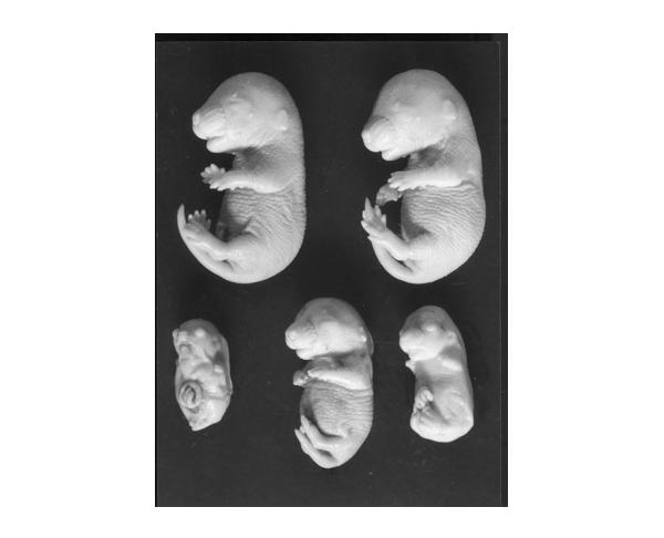
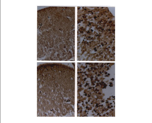
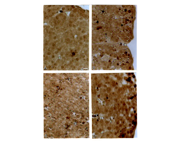
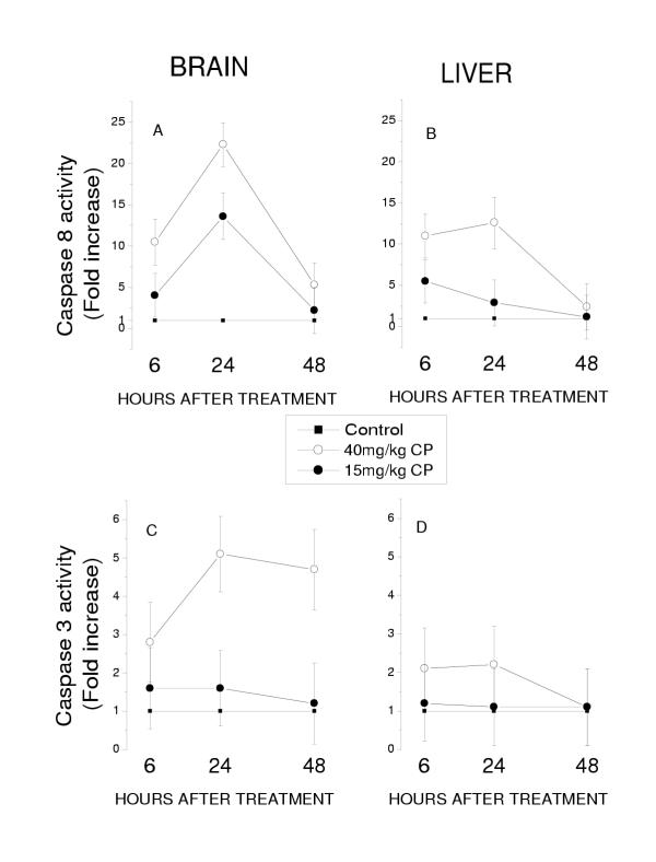
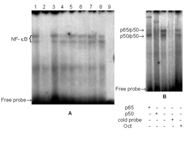
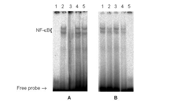
Results: The embryonic brain and liver were used as target organs. We observed that the Cyclophosphamide-induced excessive apoptosis in the brain, followed by the formation of severe craniofacial structural anomalies, was accompanied by suppression of NF-kappaB DNA-binding activity as well as by a significant and lasting increase in the activity of caspases 3 and 8. However, in the liver, in which cyclophosphamide induced transient apoptosis was not followed by dysmorphogenesis, no suppression of NF-kappaB DNA-binding activity was registered and the level of active caspases 3 and 8 was significantly lower than in the brain. It has also been observed that both the brain and liver became much more sensitive to the CP-induced teratogenic insult if the embryos were exposed to a combined treatment with the teratogen and sodium salicylate that suppressed NF-kappaB DNA-binding activity in these organs.

Conclusion: The results of this study demonstrate that suppression of NF-kappaB DNA-binding activity in embryos responding to the teratogenic insult may be associated with their decreased resistance to this insult. They also suggest that teratogens may suppress NF-kappaB DNA-binding activity in the embryonic tissues in an organ type- and dose-dependent fashion.


Figures

Figure 1
Figure 1
External structural anomalies in 17-day fetuses treated with CP alone or in combination with NaSAL. Top row: Left – a control fetus. Right – a fetus treated with NaSAL. No external anomalies were observed in these embryos. Bottom row: Left – a fetus treated with 40 mg/kg CP exhibits severe growth retardation, eventration of the abdominal wall, phocomelia and amelia (hindlimbs), absence of tail, agnathia, open eyes, severe microcephaly. Center: a fetus treated with 15 mg/kg CP exhibits light growth retardation, bowed tail, syndactyly and ectrodactyly. Right: a fetus treated with 15 mg/kg CP and NaSAL exhibits more severe growth retardation than the embryo treated with this dose of CP alone and such anomalies as microcephaly, open eyes, micrognathia, adactyly (forelimbs) and meromelia (hindlimbs), short tail, eventration of the abdominal wall.
Figure 2
Figure 2
Representative TUNEL-stained sections of the brain of E14. All sections are midsagittal and present a region of the embryonic telencephalon containing the ependyma layer, mantle and marginal layers. A – a control embryo; B – an embryo treated with 40 mg/kg CP; C – an embryo treated with NaSAL; D – an embryo treated with 15 mg/kg CP +NaSAL. Scale bar = 10 μm
Figure 3
Figure 3
Representative TUNEL-stained sections of the liver of E14. All sections are midsagittal. A – a control embryo; B – an embryo treated with 40 mg/kg CP; C – an embryo treated with NaSAL; D – an embryo treated with 40 mg/kg CP +NaSAL. Scale bar = 10 μm
Figure 4
Figure 4
Active caspase 8 and caspase 3 expression in the embryonic brain and liver of control embryos and embryos exposed to CP. Results obtained in experimental groups are presented as 95% limits for the means (fold increase) calculated by GT2-method. Means with limits that do not overlap are significantly different. Means with lower limits < 1 (the level of the activity of the caspases in controls) do not differ significantly from controls.
Figure 5
Figure 5
NF-κB complex formation in the embryonic brain and liver.A: Nuclear extracts from the brain (lanes 1–4) and liver (lanes 5–8) of E13 and E 14, control and exposed to 40 mg/kg CP. 1 – E13, control; 2 – E13, CP; 3 – E14, control; 4 – E14, CP; 5 – E13, control; 6 – E13, CP; 7 – E14, control; 8 – E14, CP; 9 – no sample.B: Supershift analysis of -κB-binding proteins presented in nuclear extract from the brain of E13 demonstrating p65/p50 heterodimers (top band) and p50/p50 homodimers (bottom band). Specificity of binding was determined by competition with 100-fold excess of unlabeled NF-κB oligonucleotide or 100-fold excess of OCT oligonucleotide.
Figure 6
Figure 6
NF-κB complex formation in the liver and brain of E14 embryos exposed CP in combination with NaSAL.A: Nuclear extracts from the liver: 1 – no sample; 2 – E14, control; 3 – E14, 40 mg/kg CP+NaSAL; 4 – E14, 40 mg/kg CP; 5 – E14, NaSAL.B: Nuclear extracts from the brain: 1 – E14, 15 mg/kg CP; 2 – E14, 15 mg/kg CP + NaSAL; 3 – E14, control; 4 – E14, 40 mg/kg CP; 5 – no sample

References

    1. Jacobson MD, Weil M, Raff MC. Programmed cell death in animal development. Cell. 1997;88:347–354. - PubMed
    1. Sadler TW, Hunter ES., III . Principles of abnormal development. Past, present and future. In: Kimmel CA, Buelke-Sam J, editor. Developmental Toxicology. New York, Raven Press; 1994. pp. 53–63.
    1. Knudsen TB. Cell death. In: Kavlock RJ, Daston GP, editor. Drug Toxicity in Embryonic Development I. Berlin, Heidelberg, Springer-Verlag; 1997. pp. 211–244.
    1. Green DR. Apoptotic pathways: the roads to ruin. Cell. 1998;94:695–698. - PubMed
    1. M. Barkett, Gilmore TD. Control of apoptosis by Rel/NF-kappaB transcription factors. Oncogene. 1999;18:6910–6924. doi: 10.1038/sj/onc/1203238. - DOI - PubMed

Publication types

MeSH terms

LinkOut - more resources