. 2002 Apr;70(4):1677-83.
doi: 10.1128/IAI.70.4.1677-1683.2002.

Peripheral blood mononuclear cell activation induced by Leptospira interrogans glycolipoprotein

Affiliations

Peripheral blood mononuclear cell activation induced by Leptospira interrogans glycolipoprotein

Decio Diament et al. Infect Immun. 2002 Apr.

Abstract

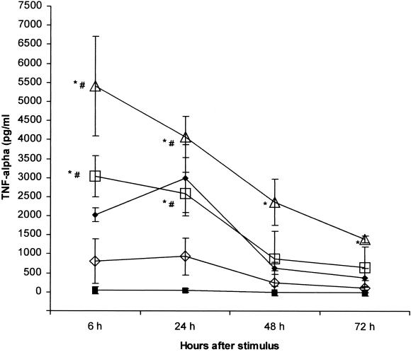
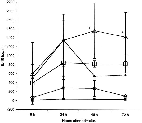
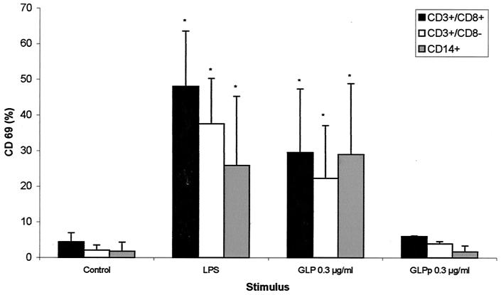
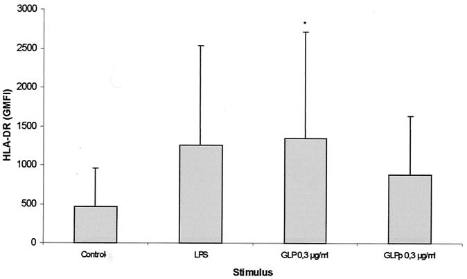
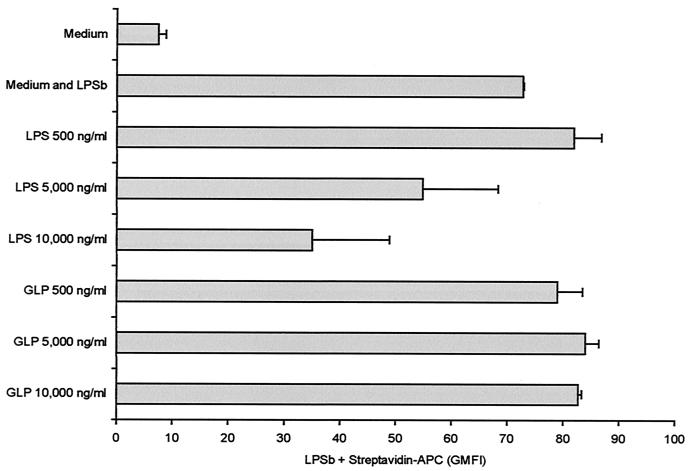
Leptospira interrogans glycolipoprotein (GLP) has been implicated in pathological and functional derangement seen in leptospirosis. The goal of this study was to evaluate GLP's ability to induce cellular activation, as assessed by cytokine production and expression of surface activation markers. GLP extracted from either pathogenic L. interrogans serovar Copenhageni or nonpathogenic Leptospira biflexa serovar Patoc (GLPp) was used to stimulate peripheral blood mononuclear cell cultures from healthy donors. Supernatant cytokine levels were measured by enzyme-linked immunosorbent assay. Expression of CD69 and HLA-DR on lymphocytes and monocytes, as well as lipopolysaccharide (LPS) binding, were measured by flow cytometry. At 6 h of incubation, GLP induced a significant rise in tumor necrosis factor alpha levels, which dropped progressively until 72 h of incubation. Interleukin-10 peak levels were obtained at between 24 and 48 h, with sustained levels until 72 h of incubation. The response magnitude was proportional to the GLP dose. CD69 expression on T lymphocytes and monocytes increased significantly, as did HLA-DR expression on monocytes. GLPp induced no CD69 or HLA-DR expression. GLP did not block biotinylated LPS binding to monocytes, suggesting that different pathways are used to induce cell activation. In conclusion, GLP induces cellular activation and may play a major role in the pathogenesis of leptospirosis.


Figures

FIG. 1.
FIG. 1.
TNF-α kinetics and dose response after stimulation with GLP. LPS was used as positive control. PBMC were stimulated with either S. enterica serovar Abortus Equi LPS or L. interrogans serovar Copenhageni GLP at the indicated doses and incubated for 72 h. Supernatants were collected at the specified time-points and TNF-α was measured by ELISA. Results are means ± SDs from three experiments. Closed squares, control; closed diamonds, LPS at 1 μg/ml; open triangles, GLP at 0.3 μg/ml; open squares, GLP at 0.03 μg/ml; open diamonds, TLP at 0.003 μg/ml. For all GLP stimuli compared with controls, P = 0.02; ∗, P < 0.05 compared to control; #, P < 0.05 compared to 0.003 μg/ml.
FIG. 2.
FIG. 2.
IL-10 kinetics and dose response after stimulation with GLP. LPS was used as positive control. PBMC were stimulated with either S. enterica serovar Abortus Equi LPS or L. interrogans serovar Copenhageni GLP in the indicated doses and incubated for 72 h. Supernatants were collected at the specified time points, and TNF-α was measured by ELISA. Results are means ± SDs from two experiments. Closed squares. control; closed diamonds, LPS at 1 μg/ml; open triangles, GLP at 0.3 μg/ml; open squares, GLP at 0.03 μg/ml; open diamonds, GLP at 0.003 μg/ml. For all GLP stimuli compared with controls, P = 0.05; ∗, P < 0.05 compared to control.
FIG. 3.
FIG. 3.
CD69 expression on LPS- or GLP-stimulated lymphocytes and monocytes. Whole blood was incubated in the presence of LPS, GLP, or medium alone for 6 h as described in Materials and Methods. CD69 is depicted as the percentage of positive cells acquired on the gate. Results are means ± SDs from five experiments with GLP and two experiments with GLPp. ∗, P < 0.05 compared to controls.
FIG. 4.
FIG. 4.
HLA-DR expression on LPS- or GLP-stimulated monocytes. Whole blood was incubated in the presence of LPS, GLP, or medium alone for 6 h as described in Materials and Methods. HLA-DR is depicted as geometric mean fluorescence intensity (GMFI). Results are means ± SDs from five experiments for GLP and two experiments for GLPp. ∗, P < 0.05 compared to controls.
FIG. 5.
FIG. 5.
LPSb binding blockade by LPS or GLP. PBMC were preincubated with increasing concentrations of LPS or GLP and then incubated with 5,000 ng of LPSb per ml. There was a decrease in the LPSb binding to PBMC preincubated with 5,000 and 10,000 ng of LPS per ml. GLP produced no blocking effect on LPSb binding to monocytes. Results are means ± SDs from two experiments. Streptavidin-APC is depicted as the geometric mean fluorescence intensity (GMFI).

Similar articles

Cited by

References

    1. Abdulkader, R. C. R., A. C. Seguro, P. S. Malheiro, E. A. Burdmann, and M. Marcondes. 1996. Peculiar electrolytic and hormonal abnormalities in acute renal failure due to leptospirosis. Am. J. Trop. Med. Hyg. 54:1-6. - PubMed
    1. Aliprantis, A. O., R. B. Yang, M. R. Mark, S. Suggett, B. Devaux, J. D. Radolf, G. R. Klimpel, P. Godowski, and A. Zychlinsky. 1999. Cell activation and apoptosis by bacterial lipoproteins through toll-like receptor-2. Science 285:736-739. - PubMed
    1. Alves, V. A. F., L. C. C. Gayotto, P. H. Yasuda, A. Wakamatsu, C. T. Kanamura, and T. Brito. 1991. Leptospiral antigens (L. interrogans serogroup ictero-haemorrhagiae) in the kidney of experimentally infected guinea pigs and their relation to the pathogenesis of the renal injury. Exp. Pathol. 42:81-93. - PubMed
    1. Barnett, J. K., D. Barnett, C. A. Bolin, T. A. Summers, E. A. Wagar, N. F. Cheville, R. A. Hartskeerl, and D. A. Haake. 1999. Expression and distribution of leptospiral outer membrane components during renal infection of hamsters. Infect. Immun. 67:853-861. - PMC - PubMed
    1. Brito, T., G. M. Bohm, and P. H. Yasuda. 1979. Vascular damage in acute experimental leptospirosis of the guinea-pig. J. Pathol. 128:177-181. - PubMed

Publication types

MeSH terms

LinkOut - more resources