. 2002 Apr;70(4):1750-60.
doi: 10.1128/IAI.70.4.1750-1760.2002.

Toxoplasma gondii-infected human myeloid dendritic cells induce T-lymphocyte dysfunction and contact-dependent apoptosis

Affiliations

Toxoplasma gondii-infected human myeloid dendritic cells induce T-lymphocyte dysfunction and contact-dependent apoptosis

Shuang Wei et al. Infect Immun. 2002 Apr.

Abstract

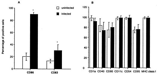
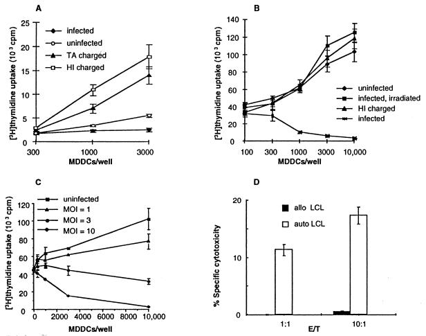
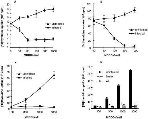
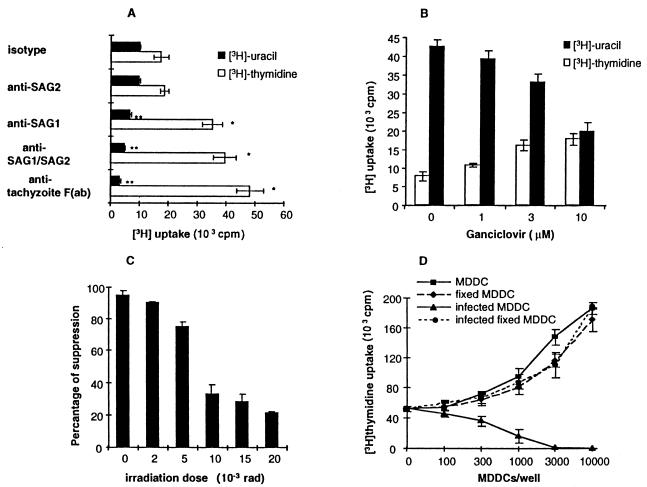
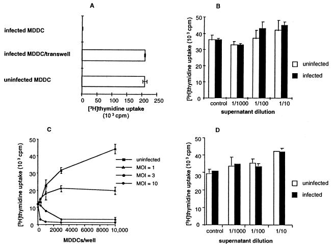
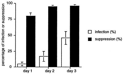
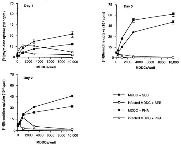
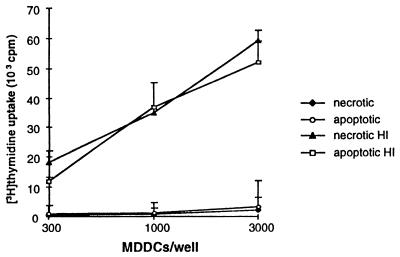
Dendritic cells ignite adaptive immunity by priming naïve T lymphocytes. Human monocyte-derived dendritic cells (MDDCs) infected with Toxoplasma gondii induce T-lymphocyte gamma interferon production and may thus activate T. gondii-specific immunity. However, we now demonstrate that T. gondii-infected MDDCs are poor at activating T lymphocytes and are unable to induce specific cytotoxic T lymphocytes. On the other hand, MDDCs acquiring nonviable T. gondii antigens directly, or indirectly through captured apoptotic or necrotic cell bodies, induce potent T-lymphocyte activation. T lymphocytes exposed to infected MDDCs are significantly impaired in upregulation of CD69 and CD28, are refractory to activation, and die through contact-dependent apoptosis mediated by an as-yet-unidentified mechanism not requiring Fas, tumor necrosis factor-related apoptosis-inducing ligand, leukocyte function antigen 1, intercellular adhesion molecule 1, tumor necrosis factor alpha, interleukin 10, alpha interferon, gamma interferon, prostaglandins, or reactive nitrogen intermediates. Bystander T lymphocytes that were neither infected nor apoptotic were refractory to activation, suggesting global dysfunction. Immunosuppression and T-lymphocyte unresponsiveness and apoptosis are typical of acute T. gondii infection. Our data suggest that infected dendritic cells contribute to these processes. On the other hand, host cells infected with T. gondii are resistant to multiple inducers of apoptosis. Thus, regulation of host cell and bystander cell apoptosis by viable T. gondii may be significant components of a strategy to evade immunity and enhance intracellular parasite survival.


Figures

FIG. 1.
FIG. 1.
T. gondii infection induces a partially mature MDDC phenotype. (A) CD86 and CD83 expressions were significantly increased after infection, and the mean relative fluorescence intensity of DR was significantly increased (see text). (B) Expression of other markers was not affected. Immature MDDCs were infected at an MOI of 10 and evaluated after 16 h. FACS analyses are of 10 individual experiments from 10 separate subjects, with the means ± SEMs shown. ∗, P < 0.01.
FIG. 2.
FIG. 2.
MDDCs infected with live T. gondii tachyzoites are defective in activating T lymphocytes. (A) T. gondii-specific T-lymphocyte activation. MDDCs infected with live tachyzoites (MOI = 10) fail to induce T-lymphocyte proliferation from seropositive subjects (P < 0.01 versus heat-inactivated [HI] tachyzoites or T. gondii Ag [TA], whereas MDDCs pulsed with 1 μg of nonviable soluble T. gondii Ag per ml or with 10 whole, heat-inactivated tachyzoites/MDDC elicited potent T-lymphocyte proliferation. (B) Killed or irradiated tachyzoites do not inhibit superantigen-mediated T-lymphocyte activation. MDDCs were pulsed with three heat inactivated tachyzoites/MDDC, or infected at an MOI of 3, or infected and irradiated with 20,000 rads from a γ source 16 h after infection. (C) Infected MDDCs inhibit superantigen-induced T-lymphocyte proliferation in a dose-dependent fashion. Peripheral blood mononuclear cells were incubated with autologous MDDCs plus 10 ng of superantigen per ml for 3 days. (D) MDDCs charged with heat-killed tachyzoites elicit T. gondii-specific cytotoxic T lymphocytes. Peripheral blood mononuclear cells from a seropositive subject were cultured with one charged MDDC per 100 peripheral blood mononuclear cells for 10 days. Cytotoxicity was assessed by 51Cr release following a 4-h incubation with autologous (auto) or allogeneic (allo) Epstein-Barr virus-transformed B cells (LCL) infected with T. gondii. The data in each panel reflect one experiment representative of five or more, except for panel D, which is representative of three experiments.
FIG. 3.
FIG. 3.
T. gondii-infected MDDCs inhibit T-lymphocyte proliferation in response to a variety of stimuli. MDDCs were infected with T. gondii strain RH (MOI = 3) and then incubated with naïve, autologous CD4+ T lymphocytes and 4 limiting flocculation units (LFU) of tetanus toxoid per ml (A) or 0.05 μg of anti-CD3 antibody per ml (B), or added to allogeneic T lymphocytes (C). (D) MDDCs infected with either A2 or Me49 T. gondii tachyzoites (MOI = 3) were defective in eliciting an allogeneic mixed lymphocyte response. Similar results with Me49- and A2-infected MDDCs were obtained with phytohemagglutinin and superantigen activation. Data shown are mean values of triplicate determinations, representative of more than five experiments for each condition, except for panel D (three experiments).
FIG. 4.
FIG. 4.
Immunosuppression mediated by T. gondii-infected MDDCs correlates directly with parasite burden and parasite replication. (A) Blockade of T. gondii attachment ligands reverses immunosuppression mediated by infected MDDCs. However, this reversal relates to reduced MDDC parasitism. MDDCs infected with tachyzoites incubated with α-SAG-1, or anti-tachyzoite F(ab) were less immunosuppressive than those infected with control antibody. Tachyzoites were incubated with antibodies shown, and then used to infect MDDCs at an MOI of 10. Superantigen-mediated activation of autologous T lymphocytes with these MDDCs was determined 3 days later. ∗ and ∗∗, P < 0.05 compared to isotype control. (B) Reduced parasite burden following infection was accomplished by using CTK11 mutant tachyzoites that are sensitive to ganciclovir (38). MDDCs were infected at an MOI of 10. Infected MDDCs were treated with ganciclovir 4 h after infection and incubated with autologous T lymphocytes in a 1:100 ratio with 10 ng of superantigen per ml in the continuous presence of drug starting 16 h after infection. Radiolabel incorporation was determined at the end of the 3-day coculture. Uracil uptake quantifies parasite replication (26). Black bars show [3H]uracil uptake by tachyzoites, and white bars show [3H]thymidine uptake by T lymphocytes. (C) Gamma irradiation of infected MDDCs 16 h following infection (MOI = 10) reduces immunosuppression. MDDCs were infected at an MOI of 10 and irradiated 16 h after infection. Superantigen-mediated activation of autologous T lymphocytes was determined as described in Materials and Methods. Percentage of suppression was calculated as follows: [3H]methylthymidine uptake of T cells exposed to infected MDDC divided by [3H]methylthymidine uptake of T cells exposed to uninfected MDDC, multiplied by 100%. (D) Paraformaldehyde-fixed, infected MDDCs are not immunosuppressive. Fixed MDDCs were prepared 14 to 18 h after infection by incubation in 0.1% paraformaldehyde at ambient temperature for 1 h, followed by thorough washing. Superantigen-mediated activation of autologous T lymphocytes was determined as described for panel B. One experiment representative of four to six is shown for panels A to D.
FIG. 5.
FIG. 5.
Direct cell-to-cell contact is required for immunosuppression mediated by T. gondii-infected MDDCs. Bystander cells do not appear to contribute to this process. (A) Peripheral blood mononuclear cells (100,000 in the upper chamber) exposed to infected MDDCs (MOI = 10; 30,000 in the lower chamber) across a semipermeable membrane of a transwell proliferated normally in response to superantigen, but not when the peripheral blood mononuclear cells and infected MDDCs were in direct contact. (B) Supernatants from purified CD1a+ MDDCs infected with T. gondii do not inhibit superantigen-mediated T-lymphocyte proliferation. Supernatant was added to a fresh, uninfected coculture. (C) T. gondii-infected, purified MDDCs (>99% CD1a+ cells) suppressed superantigen-induced proliferation of purified CD3+ T lymphocytes (>98% CD3+ cells). These experiments were set up as for Fig. 2C. (D) Supernatants from purified CD1a+ MDDCs infected with T. gondii and cocultured with T lymphocytes do not inhibit superantigen-mediated T-lymphocyte proliferation. Supernatants collected after a 3-day coculture were added to fresh, uninfected cocultures. One experiment representative of three to six is shown for all panels.
FIG. 6.
FIG. 6.
T-lymphocyte immunosuppression induced by T. gondii-infected MDDCs precedes detectable T-lymphocyte infection. MDDCs were infected with T. gondii at an MOI of 10, and infection was determined by inspection of Giemsa-stained cytospin preparations with light microscopy. At least 200 T lymphocytes per condition were analyzed. Percentage suppression was determined by dividing [3H]methylthymidine incorporation induced by T. gondii-infected MDDCs by [3H]methylthymidine incorporation induced by uninfected MDDCs, both bearing superantigen, and subtracting from 100%. Incorporation experiments were set up as described in the text using 10,000 MDDCs and 70,000 CD3+ T lymphocytes/well, and harvested on the days of culture shown. Means ± SEMs of three independent experiments using three separate subjects are reported.
FIG. 7.
FIG. 7.
Immunosuppression mediated by T. gondii-infected MDDCs appears as early as after 1 day of coculture, when only a minority of T lymphocytes are infected, and is maximal around day 2. MDDCs were infected with T. gondii at an MOI of 10 and cocultured with purified CD3+ T lymphocytes 16 h later with 10 ng of superantigen (SEB) per ml or 1 μg of phytohemagglutinin (PHA) per ml. Means ± SEMs of three independent experiments using three separate subjects are reported. See also Fig. 6.
FIG. 8.
FIG. 8.
MDDCs acquiring T. gondii antigens from necrotic or apoptotic monocytes induce T. gondii-specific T-lymphocyte proliferation. Monocytes were incubated with heat-inactivated (HI) T. gondii tachyzoites and induced to become necrotic (heat shock) or apoptotic (beauvericin treatment). Cell bodies were loaded onto autologous MDDCs, which were then incubated with autologous CD3+ T lymphocytes at a 1:10 ratio for 5 days, after which time incorporated [3H]methylthymidine incorporation was measured. Means ± SEMs of one experiment representative of four are shown. Necrotic HI, autologous monocytes loaded with heat-inactivated T. gondii tachyzoites and induced to become necrotic, etc. Necrosis versus apoptosis was confirmed by FACS for 7-AAD and propidium iodide as described in Materials and Methods.

Similar articles

Cited by

References

    1. Banchereau, J., and R. M. Steinman. 1998. Dendritic cells and the control of immunity. Nature 392:245-252. - PubMed
    1. Berens, R. L., E. C. Krug, P. B. Nash, and T. J. Curiel. 1998. Selection and characterization of Toxoplasma gondii mutants resistant to artemisinin. J. Infect. Dis. 177:1128-1131. - PubMed
    1. Brinkman, K., S. Debast, R. Sauerwein, F. Ooyman, J. Hiel, and J. Raemaekers. 1998. Toxoplasma retinitis/encephalitis 9 months after allogeneic bone marrow transplantation. Bone Marrow Transplant. 21:635-636. - PubMed
    1. Candolfi, E., C. A. Hunter, and J. S. Remington. 1994. Mitogen- and antigen-specific proliferation of T cells in murine toxoplasmosis is inhibited by reactive nitrogen intermediates. Infect. Immun. 62:1995-2001. - PMC - PubMed
    1. Candolfi, E., C. A. Hunter, and J. S. Remington. 1995. Roles of gamma interferon and other cytokines in suppression of the spleen cell proliferative response to concanavalin A and toxoplasma antigen during acute toxoplasmosis. Infect. Immun. 63:751-756. - PMC - PubMed

Publication types