. 2002 Apr;70(4):1896-908.
doi: 10.1128/IAI.70.4.1896-1908.2002.

The nucleotide sequence of Shiga toxin (Stx) 2e-encoding phage phiP27 is not related to other Stx phage genomes, but the modular genetic structure is conserved

Affiliations

The nucleotide sequence of Shiga toxin (Stx) 2e-encoding phage phiP27 is not related to other Stx phage genomes, but the modular genetic structure is conserved

Jürgen Recktenwald et al. Infect Immun. 2002 Apr.

Erratum in

  • Infect Immun 2002 Aug;70(8):4755

Abstract

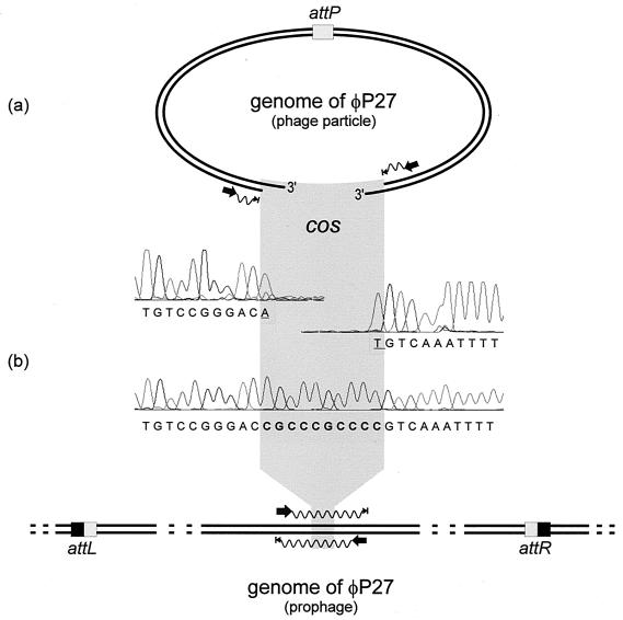
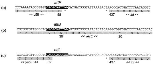
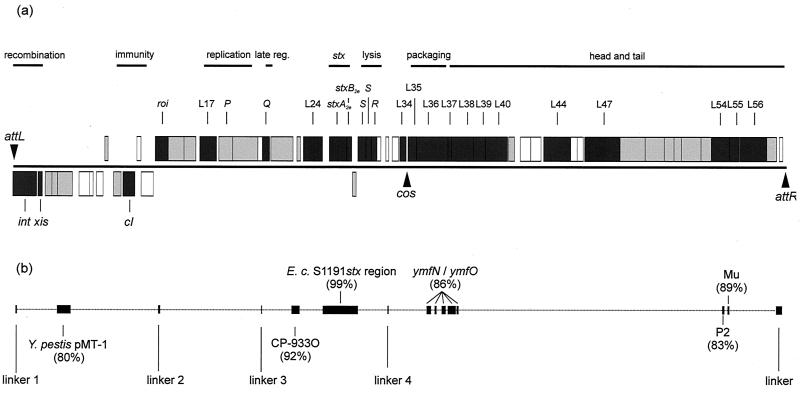
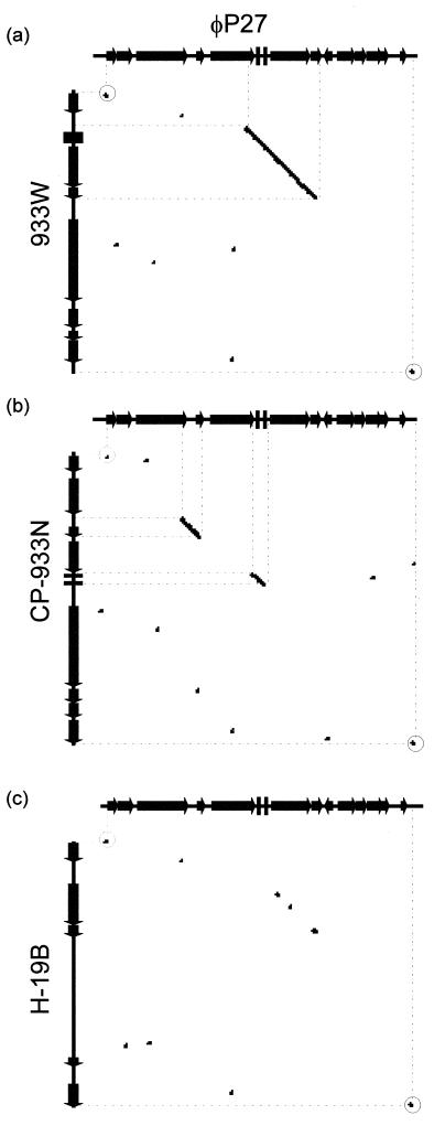
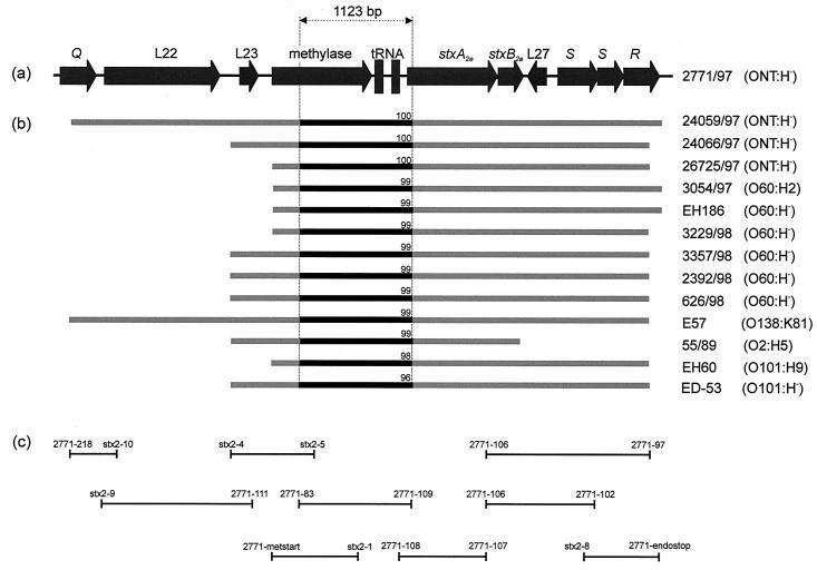
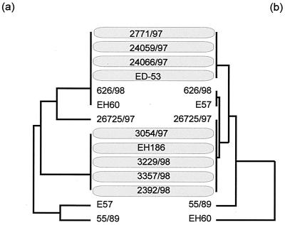
In this study we determined the complete nucleotide sequence of Shiga toxin 2e-encoding bacteriophage phi P27, isolated from the Shiga toxin-producing Escherichia coli patient isolate 2771/97. phi P27 is integrated as a prophage in the chromosomal yecE gene. This integration generates identity segments of attL and attR sites with lengths of 11 nucleotides. The integrated prophage genome has a size of 42,575 bp. We identified 58 open reading frames (ORFs), each with a length of >150 nucleotides. The deduced proteins of 44 ORFs showed significant homologies to other proteins present in sequence databases, whereas 14 putative proteins did not. For 29 proteins, we could deduce a putative function. Most of these are related to the basic phage propagation cycle. The phi P27 genome represents a mosaic composed of genetic elements which are obviously derived from related and unrelated phages. We identified five short linker sequences of 22 to 151 bp in the phi P27 sequence which have also been detected in a couple of other lambdoid phages. These linkers are located between functional modules in the phage genome and are thought to play a role in genetic recombination. Although the overall DNA sequence of phi P27 is not highly related to other known phages, the data obtained demonstrate a typical lambdoid genome structure.


Figures

FIG. 1.
FIG. 1.
Scheme of the sequencing procedure used for the identification of cos sites in the φP27 genome. (a) Sequencing primer binding sites were selected close to the ends of the linear φP27 genome to obtain runoff sequences. Electropherograms of the runoff sequences are presented. (b) In the upper part, the DNA sequence of the corresponding region sequenced from the prophage is shown. The sequencing procedure is explained in the lower part. Black arrows indicate binding sites and the directions of primers. Waves depict cycle sequence reactions that have been performed directly from the phage DNA template (a) and from a PCR product (b). Arrowheads with bars indicate the stops of sequencing reactions. The phage attachment site (attP) and the attL and attR sites generated by insertion of the phage are highlighted by boxes. Underlined nucleotides were added by the Taq polymerase independent of the template.
FIG. 2.
FIG. 2.
Determination of the phage integration site. (a) The attP site of φP27 is depicted, including parts of ORF L58 and int. (b) The corresponding attB site in the chromosomal yecE gene is shown. (c) Depiction of the DNA sequence of one of the borders of the integrated prophage. According to WebGeneMark.hmm, integration of the prophage into yecE leads to a truncated protein. The upper lines are the nucleotide sequences, and the lower lines are the amino acid sequences. The numbers below the amino acid sequence mark the amino acid positions of the respective protein. yecE′ labels the truncated amino acid sequence, asterisks indicate stop codons, and arrows indicate the transcription orientation. Black boxes in the nucleotide sequences indicate the identity segment of attachment sites.
FIG. 3.
FIG. 3.
Schematic illustration of the φP27 genome. (a) Boxes depict ORFs determined by WebGenemark.hmm. ORFs directed in rightward orientation are drawn above the black line; ORFs directed in the leftward direction are drawn below the line. Boxes in dark gray represent ORFs encoding proteins described in Results. Boxes in light gray mark ORFs, the deduced proteins of which show homology to known proteins which are described only in Table 1. Empty boxes label ORFs encoding proteins without known homologies. (b) Black boxes and vertical bars represent regions with nucleotide sequence homologies to other sequences, including linkers. Homology values are depicted by boxes. late reg., late regulation.
FIG. 4.
FIG. 4.
Dot blot matrix of the stx2e-flanking region of φP27 (genes Q to L32) and corresponding regions of phages 933W (a), CP-933N (b), and H-19B (c). At every point in the matrix where the two sequences are identical a dot is placed. A diagonal stretch of dots indicates regions where the two sequences are similar. Dots with circles represent linkers 3 (left side) and 4 (right side).
FIG. 5.
FIG. 5.
Schematic illustration of the PCR and sequencing approach used for characterization of stx2e-flanking regions of different STEC isolates. (a) Map of the stx2e-flanking region of φP27 spanning from Q to R. (b) Bars in light gray indicate the regions analyzed by PCR and initial sequencing with the same primers as employed for PCR. Black bars between dotted vertical lines depict regions which have been completely sequenced and used for the phylogenetic analysis depicted in Fig. 6. Numbers on black bars show percent DNA identity of the fragments compared to the corresponding fragment of φP27. (c) Size and location of PCR products and applied primers.
FIG. 6.
FIG. 6.
Phylogenetic analyses and comparison of sequences obtained from the 1,123-bp 5′ stx2e-flanking region and the housekeeping gene aroE from particular STEC isolates. We used GrowTree (HUSAR software package) to create a phylogenetic tree from a distance matrix created by the program by using the unweighted pair group method with arithmetic averages. The left side contains aroE sequences (a), and the right side contains phage sequences (b).

Similar articles

Cited by

References

    1. Altschul, S. F., W. Gish, W. Miller, E. W. Myers, and D. J. Lipman. 1990. Basic local alignment search tool. J. Mol. Biol. 215:403-410. - PubMed
    1. Anton, I. A., and J. R. Coggins. 1988. Sequencing and overexpression of the Escherichia coli aroE gene encoding shikimate dehydrogenase. Biochem. J. 249:319-326. - PMC - PubMed
    1. Argos, P., A. Landy, K. Abremski, J. B. Egan, E. Haggard-Ljungquist, R. H. Hoess, M. L. Kahn, B. Kalionis, S. V. Narayana, L. S. Pierson III, et al. 1986. The integrase family of site-specific recombinases: regional similarities and global diversity. EMBO J. 5:433-440. - PMC - PubMed
    1. Bateman, A., E. Birney, R. Durbin, S. R. Eddy, K. L. Howe, and E. L. Sonnhammer. 2000. The Pfam protein families database. Nucleic Acids Res. 28:263-266. - PMC - PubMed
    1. Besemer, J., and M. Borodovsky. 1999. Heuristic approach to deriving models for gene finding. Nucleic Acids Res. 27:3911-3920. - PMC - PubMed

Publication types