. 2002 Apr;70(4):1936-48.
doi: 10.1128/IAI.70.4.1936-1948.2002.

In vivo clearance of an intracellular bacterium, Francisella tularensis LVS, is dependent on the p40 subunit of interleukin-12 (IL-12) but not on IL-12 p70

Affiliations

In vivo clearance of an intracellular bacterium, Francisella tularensis LVS, is dependent on the p40 subunit of interleukin-12 (IL-12) but not on IL-12 p70

Karen L Elkins et al. Infect Immun. 2002 Apr.

Abstract

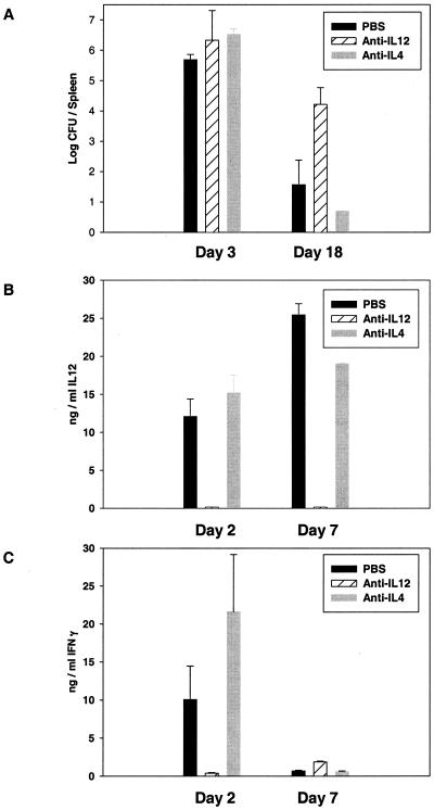
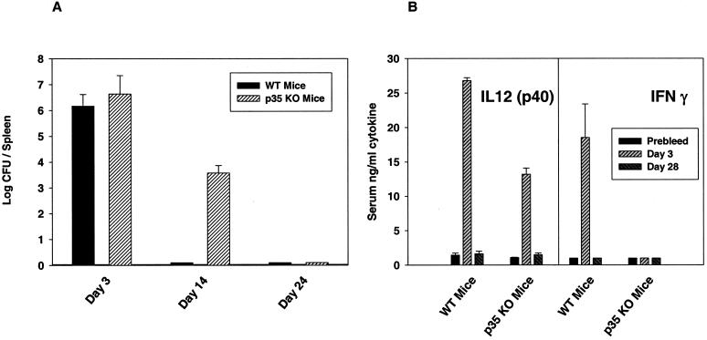
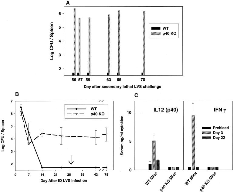
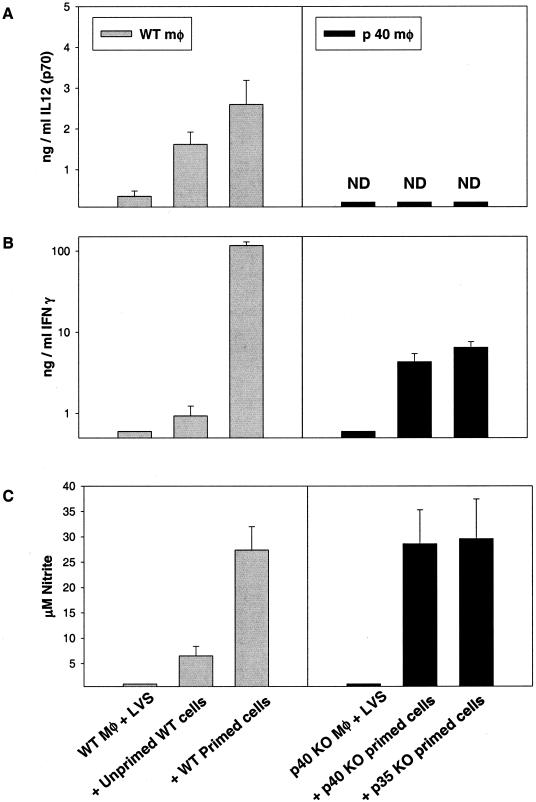
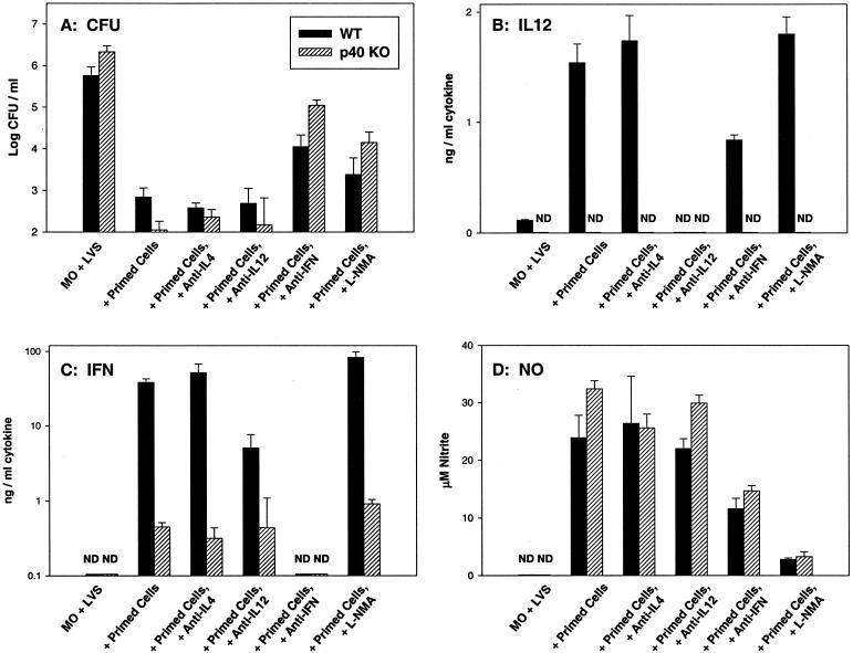
To determine the role of interleukin-12 (IL-12) in primary and secondary immunity to a model intracellular bacterium, we have comprehensively evaluated infection with Francisella tularensis LVS in three murine models of IL-12 deficiency. Mice lacking the p40 protein of IL-12 (p40 knockout [KO] mice) and mice treated in vivo with neutralizing anti-IL-12 antibodies survived large doses of primary and secondary LVS infection but never cleared bacteria and exhibited a chronic infection. In dramatic contrast, mice lacking the p35 protein (p35 KO mice) of heterodimeric IL-12 readily survived large doses of primary sublethal LVS infection as well as maximal secondary lethal challenge, with only a slight delay in clearance of bacteria. LVS-immune wild-type (WT) lymphocytes produced large amounts of gamma interferon (IFN-gamma), but p35 KO and p40 KO lymphocytes produced much less; nonetheless, similar amounts of NO were found in all cultures containing immune lymphocytes, and all immune lymphocytes were equally capable of controlling intracellular growth of LVS in vitro. Purified CD4(+) and CD8(+) T cells from both WT and p40 KO mice controlled intracellular growth, even though T cells from WT mice produced much more IFN-gamma than those from p40 KO mice, and p40 KO T cells did not adopt a Th2 phenotype. Thus, while IL-12 p70 stimulation of IFN-gamma production may be important for bacteriostasis, IL-12 p70 is not necessary for appropriate development of LVS-immune T cells that are capable of controlling intracellular bacterial growth and for clearance of primary or secondary LVS infection. Instead, an additional mechanism dependent on the IL-12 p40 protein, either alone or in another complex such as the newly discovered heterodimer IL-23, appears to be responsible for actual clearance of this intracellular bacterium.


Figures

FIG. 1.
FIG. 1.
Bacterial burdens and serum cytokine levels in anti-IL-12-treated mice following primary sublethal F. tularensis LVS infection. BALB/cByJ mice were treated with PBS or anti-cytokine antibodies as described in Table 1 and infected with 106 LVS i.d. At the indicated times after infection, groups of four mice were sacrificed for determination of bacterial burdens in spleens (A) and to obtain serum for assessment of IL-12 p40 protein (B) or IFN-γ (C) by ELISA. No IL-12 p40 or IFN-γ was detected in prebleed serum samples from any mice; the limit of detection was approximately 150 pg/ml. Results from one representative experiment of four independent experiments of similar design are shown.
FIG. 2.
FIG. 2.
Bacterial burdens and serum cytokine levels in p35 KO mice following primary sublethal F. tularensis LVS infection. C57BL/6J WT or C57-p35 KO mice were infected with 106 LVS i.d. At the indicated times after infection, groups of four mice were sacrificed for determination of bacterial burdens in spleens (A) and to obtain serum for assessment of IL-12 p40 protein or IFN-γ (B) by ELISA; the limit of detection was approximately 150 pg/ml. Results from one representative experiment of four independent experiments of similar design are shown.
FIG. 3.
FIG. 3.
Bacterial burdens and serum cytokine levels in p40 KO mice following primary and secondary F. tularensis LVS infection. BALB/cByJ WT or BALB/c-p40 KO mice were infected with 106 LVS i.d. and challenged on day 30 (arrow) with 106 LVS i.p. (1,000,000 LD50s for BALB/cByJ mice). At the indicated times after secondary lethal challenge, individual mice were sacrificed for determination of bacterial burdens in spleens (A). Other C57BL/6J WT or C57-p40 KO mice, in groups of three, were sacrificed at the indicated time points after primary or secondary (arrow) LVS infection for determination of bacterial burdens in spleens (B), or serum was obtained after primary LVS infection for assessment of IL-12 p40 protein (C, left panel) or IFN-γ (C, right panel) by ELISA; the limit of detection was approximately 150 pg/ml. Results from one representative experiment of two (B) or three (C) independent experiments of similar design are shown.
FIG. 4.
FIG. 4.
LVS-immune lymphocytes from WT, p35, or p40 KO mice control intracellular LVS growth in vitro. BMMφ were derived from either WT C57BL/6J mice (left) or from C57-p40 KO mice (right) and infected with LVS. Splenic lymphocytes from the indicated mice (unprimed C57BL/6J mice or LVS-primed mice [mice that were infected with 106 LVS i.d. on day 0 and then challenged on day 35 with 5 × 10⁶ LVS i.p., with spleens removed 4 weeks later]) were added immediately after infection, and growth of bacteria in macrophages was monitored over time. Results shown are the mean CFU/well ± standard deviations (SD) of three triplicate culture wells per condition. Results from one representative experiment of two (using lymphocytes from C57-p35 KO mice) or nine (using lymphocytes from either BALB/c or C57-p40 KO mice) independent experiments of similar design are shown.
FIG. 5.
FIG. 5.
Cytokine and NO production during in vitro control of intracellular LVS growth by WT, p35, or p40 LVS-immune lymphocytes. Supernatants were obtained at 72 h from the indicated cultures described for Fig. 4 and assessed by ELISA for the presence of IL-12 p40 protein (A) or IFN-γ (B) or by Greiss reaction for nitrite as an indicator of NO production (C). Results shown are the mean nanograms of cytokine per milliliter or micromolar NO ± SD of three triplicate culture wells per condition. Results for IFN-γ are shown on a log scale because of the large differences in quantities between the levels of C57BL/6J WT and C57-KO mice. Results from one representative experiment of two (using lymphocytes from C57-p35 KO mice) or nine (using lymphocytes from either BALB/c or C57-p40 KO mice) independent experiments of similar design are shown.
FIG. 6.
FIG. 6.
Effect of anticytokine treatment on in vitro control of intracellular LVS growth and production of mediators by WT or p40 LVS-immune lymphocytes. BMMφ were derived from either WT C57BL/6J mice or from C57-p40 KO mice and infected with LVS. Splenic lymphocytes from either C57BL/6J WT or C57-p40 KO immune mice (that had received a primary sublethal i.d. LVS infection of 105 CFU 35 days earlier) were added immediately after infection. Results using only LVS-infected WT macrophages with WT immune lymphocytes (black bars) or LVS-infected p40 KO macrophages with p40 KO immune lymphocytes (hatched bars) are shown. Growth of bacteria in macrophages was assessed at 72 h after infection (A); results shown are the mean CFU/well ± SD of three triplicate culture wells per condition. Supernatants were obtained at 72 h from the same cultures and assessed by ELISA for the presence of IL-12 p40 protein (B) or IFN-γ (C) or by Greiss reaction for nitrite as an indicator of NO production (D). Results shown are the mean nanograms of cytokine per milliliter or micromolar NO ± SD of three triplicate culture wells per condition. Results for IFN-γ are shown on a log scale because of the large differences in quantities between the levels of WT and KO mice. Results from one representative experiment of six independent experiments of similar design are shown.
FIG. 7.
FIG. 7.
In vitro control of intracellular LVS growth, cytokine production, and NO production by WT or p40 LVS-immune T-cell subpopulations. BMMφ were derived from either WT C57BL/6J mice or C57-p40 KO mice and infected with LVS. Splenic lymphocytes or purified CD4+ or CD8+ T cells from either C57BL/6J WT or C57-p40 KO immune mice (that had received a primary sublethal i.d. LVS infection of 105 CFU 45 days earlier) were added to the homologous LVS-infected macrophages immediately after infection. By flow cytometry, WT CD4+ T cells were 94.3% CD4+, WT CD8+ T cells were 90.8% CD8+, p40 KO CD4+ T cells were 90.4% CD4+, and p40 KO CD8+ T cells were 87.3% CD8+. Results using only LVS-infected WT macrophages with WT immune lymphocytes (black bars) or LVS-infected p40 KO macrophages with p40 KO immune lymphocytes (hatched bars) are shown. Growth of bacteria in macrophages was assessed at 72 h after infection (A); results shown are the mean CFU/well ± SD of three triplicate culture wells per condition. Supernatants were obtained at 72 h from the same cultures and assessed by ELISA for the presence of IL-12 p40 protein (B) or IFN-γ (C) or by Greiss reaction for nitrite as an indicator of NO production (D). Results shown are the mean nanograms of cytokine per milliliter or micromolar NO ± SD of three triplicate culture wells per condition. Results for IFN-γ are shown on a log scale because of the large differences in quantities between the levels of WT and KO mice. Results from one representative experiment of three independent experiments of similar design using WT and p40 KO cultures are shown.

Similar articles

Cited by

References

    1. Anthony, L. S. D., E. Ghadirian, F. P. Nestel, and P. A. L. Kongshavn. 1989. The requirement for gamma interferon in resistance of mice to experimental tularemia. Microb. Pathog. 7:421-428. - PubMed
    1. Badovinac, V. P., and J. T. Harty. 2000. Adaptive immunity and enhanced CD8+ T cell response to Listeria monocytogenes in the absence of perforin and IFN-gamma. J. Immunol. 164:6444-6452. - PubMed
    1. Baker, C. N., D. G. Hollis, and C. Thornsberry. 1985. Antimicrobial susceptibility testing of Francisella tularensis with a modified Mueller-Hinton broth. J. Clin. Microbiol. 22:212-215. - PMC - PubMed
    1. Bosio, C. M., and K. L. Elkins. 2001. Susceptibility to secondary Francisella tularensis LVS infection in B-cell-deficient mice is associated with neutrophilia but not with defects in specific T-cell-mediated immunity. Infect. Immun. 69:194-203. - PMC - PubMed
    1. Brombacher, F., A. Dorfmuller, J. Magram, W. J. Dai, G. Kohler, A. Wunderlin, K. Palmer-Lehmann, M. K. Gately, and G. Alber. 1999. IL-12 is dispensable for innate and adaptive immunity against low doses of Listeria monocytogenes. Int. Immunol. 11:325-332. - PubMed