

Mice lacking T and B lymphocytes develop transient colitis and crypt hyperplasia yet suffer impaired bacterial clearance during Citrobacter rodentium infection
- PMID: 11895973
- PMCID: PMC127821
- DOI: 10.1128/IAI.70.4.2070-2081.2002
Mice lacking T and B lymphocytes develop transient colitis and crypt hyperplasia yet suffer impaired bacterial clearance during Citrobacter rodentium infection
Abstract
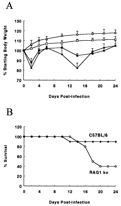
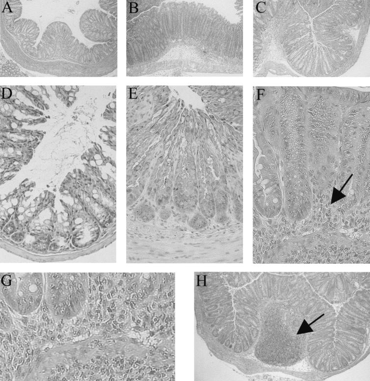
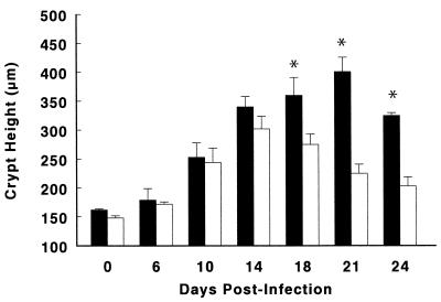
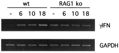
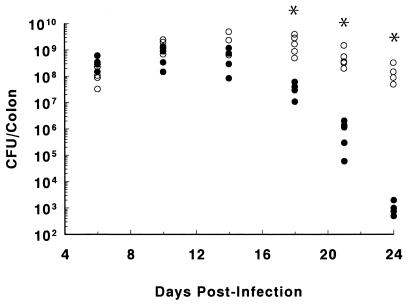
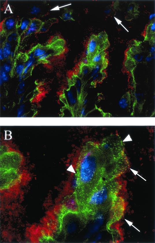
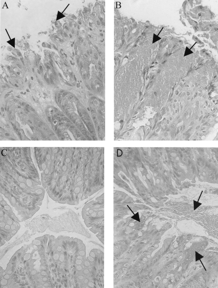
The bacterial pathogen Citrobacter rodentium belongs to a family of gastrointestinal pathogens that includes enteropathogenic and enterohemorrhagic Escherichia coli and is the causative agent of transmissible colonic hyperplasia in mice. The molecular mechanisms used by these pathogens to colonize host epithelial surfaces and form attaching and effacing (A/E) lesions have undergone intense study. In contrast, little is known about the host's immune response to these infections and its importance in tissue pathology and bacterial clearance. To address these issues, wild-type mice and mice lacking T and B lymphocytes (RAG1 knockout [KO]) were infected with C. rodentium. By day 10 postinfection (p.i.), both wild-type and RAG1 KO mice developed colitis and crypt hyperplasia, and these responses became more exaggerated in wild-type mice over the next 2 weeks, as they cleared the infection. By day 24 p.i., bacterial clearance was complete, and the colitis had subsided; however, crypt heights remained increased. In contrast, inflammatory and crypt hyperplastic responses in the RAG1 KO mice were transient, subsiding after 2 weeks. By day 24 p.i., RAG1 KO mice showed no signs of bacterial clearance and infection was often fatal. Surprisingly, despite remaining heavily infected, tissues from RAG1 KO mice surviving the acute colitis showed few signs of disease. These results thus emphasize the important contribution of the host immune response during infection by A/E bacterial pathogens. While T and/or B lymphocytes are essential for host defense against C. rodentium, they also mediate much of the tissue pathology and disease symptoms that occur during infection.
Figures
Similar articles
-
Central role for B lymphocytes and CD4+ T cells in immunity to infection by the attaching and effacing pathogen Citrobacter rodentium.Infect Immun. 2003 Sep;71(9):5077-86. doi: 10.1128/IAI.71.9.5077-5086.2003. Infect Immun. 2003. PMID: 12933850 Free PMC article.
-
Modulation of intestinal goblet cell function during infection by an attaching and effacing bacterial pathogen.Infect Immun. 2008 Feb;76(2):796-811. doi: 10.1128/IAI.00093-07. Epub 2007 Nov 5. Infect Immun. 2008. PMID: 17984203 Free PMC article.
-
Impaired resistance and enhanced pathology during infection with a noninvasive, attaching-effacing enteric bacterial pathogen, Citrobacter rodentium, in mice lacking IL-12 or IFN-gamma.J Immunol. 2002 Feb 15;168(4):1804-12. doi: 10.4049/jimmunol.168.4.1804. J Immunol. 2002. PMID: 11823513
-
Molecular pathogenesis of Citrobacter rodentium and transmissible murine colonic hyperplasia.Microbes Infect. 2001 Apr;3(4):333-40. doi: 10.1016/s1286-4579(01)01387-9. Microbes Infect. 2001. PMID: 11334751 Review.
-
Host defences to Citrobacter rodentium.Int J Med Microbiol. 2003 Apr;293(1):87-93. doi: 10.1078/1438-4221-00247. Int J Med Microbiol. 2003. PMID: 12755369 Review.
Cited by
-
TRPV1 controls innate immunity during Citrobacter rodentium enteric infection.PLoS Pathog. 2023 Dec 18;19(12):e1011576. doi: 10.1371/journal.ppat.1011576. eCollection 2023 Dec. PLoS Pathog. 2023. PMID: 38109366 Free PMC article.
-
Toll-like receptor 4 contributes to colitis development but not to host defense during Citrobacter rodentium infection in mice.Infect Immun. 2006 May;74(5):2522-36. doi: 10.1128/IAI.74.5.2522-2536.2006. Infect Immun. 2006. PMID: 16622187 Free PMC article.
-
Host engulfment pathway controls inflammation in inflammatory bowel disease.FEBS J. 2020 Sep;287(18):3967-3988. doi: 10.1111/febs.15236. Epub 2020 Feb 20. FEBS J. 2020. PMID: 32003126 Free PMC article.
-
The apolipoprotein E-mimetic peptide COG112 inhibits NF-kappaB signaling, proinflammatory cytokine expression, and disease activity in murine models of colitis.J Biol Chem. 2011 Feb 4;286(5):3839-50. doi: 10.1074/jbc.M110.176719. Epub 2010 Nov 29. J Biol Chem. 2011. PMID: 21115487 Free PMC article.
-
Indole-3-Carbinol Inhibits Citrobacter rodentium Infection through Multiple Pathways Including Reduction of Bacterial Adhesion and Enhancement of Cytotoxic T Cell Activity.Nutrients. 2020 Mar 27;12(4):917. doi: 10.3390/nu12040917. Nutrients. 2020. PMID: 32230738 Free PMC article.
References
-
- Agin, T. S., J. R. Cantey, E. C. Boedeker, and M. K. Wolf. 1996. Characterization of the eaeA gene from rabbit enteropathogenic Escherichia coli strain RDEC-1 and comparison to other eaeA genes from bacteria that cause attaching-effacing lesions. FEMS Microbiol. Lett. 144:249-258. - PubMed
-
- An, H., J. M. Fairbrother, C. Desautels, T. Mabrouk, D. Dugourd, H. Dezfulian, and J. Harel. 2000. Presence of the LEE (locus of enterocyte effacement) in pig attaching and effacing Escherichia coli and characterization of eae, espA, espB and espD genes of PEPEC (pig EPEC) strain 1390. Microb. Pathog. 28:291-300. - PubMed
-
- Artis, D., C. S. Potten, K. J. Else, F. D. Finkelman, and R. K. Grencis. 1999. Trichuris muris: host intestinal epithelial cell hyperproliferation during chronic infection is regulated by interferon-gamma. Exp. Parasitol. 92:144-153. - PubMed
-
- Bancroft, A. J., and R. K. Grencis. 1998. Th1 and Th2 cells and immunity to intestinal helminths. Chem. Immunol. 71:192-208. - PubMed
Publication types
MeSH terms
Substances
LinkOut - more resources
Full Text Sources
Other Literature Sources
Research Materials
