. 2002 Apr;70(4):2187-97.
doi: 10.1128/IAI.70.4.2187-2197.2002.

Balance of pro- and anti-inflammatory cytokines correlates with outcome of acute experimental Pseudomonas aeruginosa keratitis

Affiliations

Balance of pro- and anti-inflammatory cytokines correlates with outcome of acute experimental Pseudomonas aeruginosa keratitis

A Thakur et al. Infect Immun. 2002 Apr.

Abstract

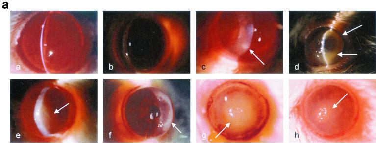
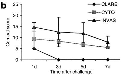
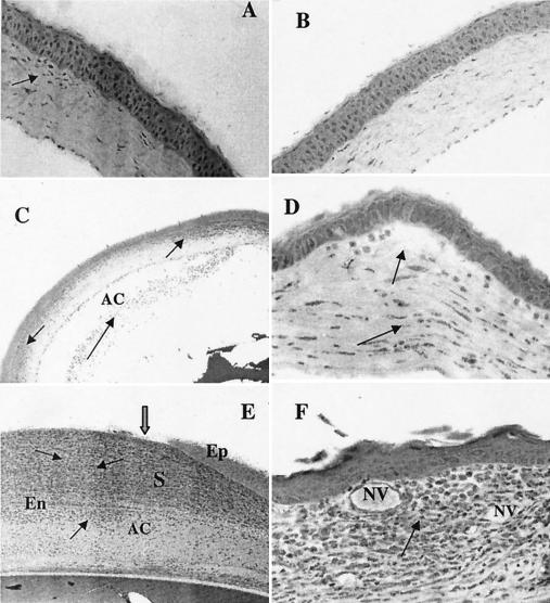
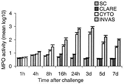
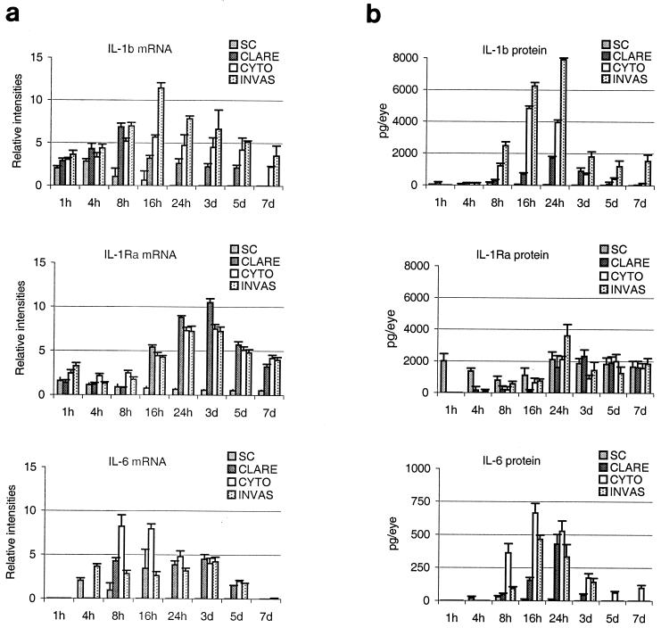
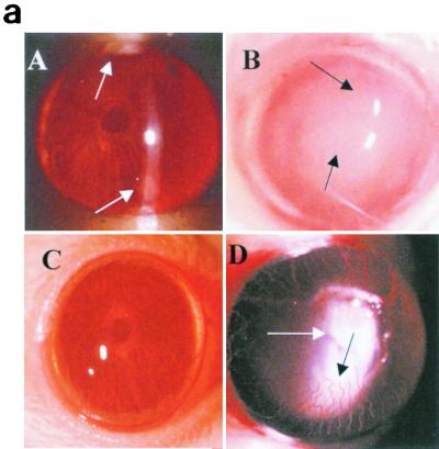
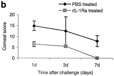
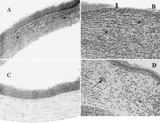
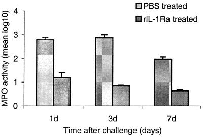
The purpose of this study was to elucidate the expression of pro- and anti-inflammatory cytokines in mouse corneas infected with Pseudomonas aeruginosa. Three bacterial strains (invasive, cytotoxic, or CLARE [contact lens-induced acute red eye]) which have recently been shown to produce distinct patterns of corneal disease in the mouse were used. The left mouse (BALB/c) corneas were scarified and infected with 2 x 10(6) CFU of one of the three P. aeruginosa strains, while right eyes served as controls. Animals were examined at 1, 4, 8, 16, and 24 h with a slit lamp biomicroscope to grade the severity of infection. Following examination, eyes were collected and processed for histopathology, multiprobe RNase protection assay for cytokine mRNA, enzyme-linked immunosorbent assay to quantitate cytokine proteins, and myeloperoxidase activity to quantitate polymorphonuclear leukocytes. The kinetics of appearance and magnitude of expression of key cytokines varied significantly in the three different phenotypes of P. aeruginosa infection. The predominant cytokines expressed in response to all three phenotypes were interleukin-1 beta (IL-1 beta), IL-1Ra, and IL-6. In response to the invasive strain, which induced severe corneal inflammation, significantly lower ratios of IL-1Ra to IL-1 beta were present at all time points, whereas corneas challenged with the CLARE strain, which induced very mild inflammation, showed a high ratio of IL-1Ra to IL-1 beta. The outcome of infection in bacterial keratitis correlated with the relative induction of these pro- and anti-inflammatory cytokines, and exogenous administration of recombinant rIL-1Ra (rIL-1Ra) was able to reduce the disease severity significantly. These findings point to the therapeutic potential of rIL-1Ra protein in possible treatment strategies for bacterial keratitis.


Figures

FIG. 1.
FIG. 1.
(a) Clinical examination of mouse corneas inoculated with the CLARE, cytotoxic, or invasive bacterial strain of P. aeruginosa. Panel a, CLARE strain at 1 day postchallenge; few focal infiltrates were present in the cornea. Panel b, CLARE strain at 7 days postchallenge; the cornea appeared normal. Panel c, cytotoxic strain at 8 h postchallenge; the cornea shows diffuse and focal infiltrates mostly in the periphery (arrow). Panel d, cytotoxic strain at 1 day postchallenge; the cornea shows dense infiltrates in the periphery, making a ring appearance (arrow). Panel e, cytotoxic strain at 7 days postchallenge; infiltrates are still present in the central cornea (arrow). Panel f, invasive strain at 8 h postchallenge; the cornea shows infiltrates extending from the limbus to the periphery (arrow). Panel g, invasive strain at 1 day postchallenge; the cornea shows dense infiltrates extending from the central cornea to the periphery (arrow). Panel h, invasive strain at: 7 days postchallenge; the corneal pathology had resolved somewhat (arrow shows infiltrates). (b) Composite clinical scores (composed of the graded density, depth, and extent of corneal infiltrates as well as size and depth of corneal ulceration; see Table 1 for the grading system) for corneas challenged with the invasive (INVAS), cytotoxic (CYTO), and CLARE bacterial strains. Error bars indicate standard errors of the means.
FIG. 1.
FIG. 1.
(a) Clinical examination of mouse corneas inoculated with the CLARE, cytotoxic, or invasive bacterial strain of P. aeruginosa. Panel a, CLARE strain at 1 day postchallenge; few focal infiltrates were present in the cornea. Panel b, CLARE strain at 7 days postchallenge; the cornea appeared normal. Panel c, cytotoxic strain at 8 h postchallenge; the cornea shows diffuse and focal infiltrates mostly in the periphery (arrow). Panel d, cytotoxic strain at 1 day postchallenge; the cornea shows dense infiltrates in the periphery, making a ring appearance (arrow). Panel e, cytotoxic strain at 7 days postchallenge; infiltrates are still present in the central cornea (arrow). Panel f, invasive strain at 8 h postchallenge; the cornea shows infiltrates extending from the limbus to the periphery (arrow). Panel g, invasive strain at 1 day postchallenge; the cornea shows dense infiltrates extending from the central cornea to the periphery (arrow). Panel h, invasive strain at: 7 days postchallenge; the corneal pathology had resolved somewhat (arrow shows infiltrates). (b) Composite clinical scores (composed of the graded density, depth, and extent of corneal infiltrates as well as size and depth of corneal ulceration; see Table 1 for the grading system) for corneas challenged with the invasive (INVAS), cytotoxic (CYTO), and CLARE bacterial strains. Error bars indicate standard errors of the means.
FIG. 2.
FIG. 2.
Histological examination of mouse corneas at 1 and 7 days postchallenge. (A) Cornea inoculated with the CLARE strain showed focal infiltrates over the anterior corneal stroma (arrow). The initial scratch site was healed completely within 24 h of challenge. (B) At 7 days postchallenge with the CLARE strain, the cornea appeared to be completely normal. (C) Corneas inoculated with the cytotoxic strain also showed massive PMN infiltration, with leukocytes streaming through limbus and conjunctiva into the periphery of the corneal stroma (arrow) and to a lesser extent in the central cornea. Infiltrating cells were also present in anterior chamber (arrow). (D) At 7 days postchallenge with the cytotoxic strain, the cornea showed infiltrates in the central region (arrow) and less so in the peripheral cornea. The epithelium had completely healed by this time point. Stromal destruction was evident in the cornea as a result of extracellular matrix destruction, appearing as large white space (arrow). (E) Corneas inoculated with the invasive bacterial strain showed massive PMN infiltration (arrows) into the central stroma at 24 h postchallenge. The epithelium in the central cornea was completely destroyed (thick arrow). Large numbers of infiltrating cells were also present in the anterior chamber (arrow). (F) At 7 days postchallenge, the corneal epithelium was healed but infiltrates were still present (arrow) and new vessel growth was evident in the mid-periphery of the cornea. Ep, epithelium; S, stroma; En, endothelium; AC, anterior chamber; NV, new blood vessels.
FIG. 3.
FIG. 3.
MPO activity in whole eyes challenged with the invasive (INVAS), cytotoxic (CYTO), or CLARE strain and in a scratch control (SC) at 1, 4, 8, 16, and 24 h and 3, 5, and 7 days postchallenge. Results are reported as mean (± standard error of the mean) log10 MPO activity/eye. Note that the horizontal axis is not on a linear scale.
FIG. 4.
FIG. 4.
(a) Time-dependent expression of cytokine mRNAs in mouse corneas at 1, 4, 8, 16, and 24 h and 3, 5, and 7 days postchallenge with the invasive (INVAS), cytotoxic (CYTO), or CLARE bacterial strain. Results are presented as the mean (± standard error of the mean) relative intensity of each cytokine band divided by the intensity of the respective GAPDH band in the same lane. (b) Time dependent expression of cytokine proteins in mouse corneas at 1, 4, 8, 16, and 24 h and 3, 5, and 7 days postchallenge with the invasive, cytotoxic, or CLARE bacterial strain. Results are presented as mean (± standard error of the mean) picograms of cytokine protein/eye. Note that the horizontal axis is not on a linear scale. SC, scratch control.
FIG. 5.
FIG. 5.
(a) Clinical examination of corneas injected with 20 μl of rIL-1Ra (20 μg during each injection) subconjunctivally at 24 h and then 3 h before infection with the invasive strain. Control mice received an equal volume of PBS at same time points before the infection with invasive strain. Panel A, rIL-1Ra-treated mice at 24 h postchallenge, showing focal infiltrates at the periphery of the cornea (arrows). Panel B, control mice showed extensive infiltration of inflammatory cells (arrow) in the central cornea at 24 h postchallenge, with severe edema and moderate anterior chamber response. Panel C, rIL-1Ra-treated mice at 7 days postchallenge; infiltrates have completely resolved. Panel D, in control mice the epithelium had healed but a large number of infiltrates were still present (white arrow), and new vessel growth was evident (black arrow). (b) Composite clinical scores (composed of the graded density, depth, and extent of corneal infiltrates as well as size and depth of corneal ulceration; see Table 1 for the grading system) at 1, 3, and 7 days postchallenge for corneas treated with 20 μl of rIL-1Ra (20 μg during each injection) subconjunctivally at 24 and 3 h before the infection with invasive strain. Control mice received an equal volume of PBS at same time points before the infection with invasive strain. Error bars indicate standard errors of the means.
FIG. 5.
FIG. 5.
(a) Clinical examination of corneas injected with 20 μl of rIL-1Ra (20 μg during each injection) subconjunctivally at 24 h and then 3 h before infection with the invasive strain. Control mice received an equal volume of PBS at same time points before the infection with invasive strain. Panel A, rIL-1Ra-treated mice at 24 h postchallenge, showing focal infiltrates at the periphery of the cornea (arrows). Panel B, control mice showed extensive infiltration of inflammatory cells (arrow) in the central cornea at 24 h postchallenge, with severe edema and moderate anterior chamber response. Panel C, rIL-1Ra-treated mice at 7 days postchallenge; infiltrates have completely resolved. Panel D, in control mice the epithelium had healed but a large number of infiltrates were still present (white arrow), and new vessel growth was evident (black arrow). (b) Composite clinical scores (composed of the graded density, depth, and extent of corneal infiltrates as well as size and depth of corneal ulceration; see Table 1 for the grading system) at 1, 3, and 7 days postchallenge for corneas treated with 20 μl of rIL-1Ra (20 μg during each injection) subconjunctivally at 24 and 3 h before the infection with invasive strain. Control mice received an equal volume of PBS at same time points before the infection with invasive strain. Error bars indicate standard errors of the means.
FIG. 6.
FIG. 6.
Animals were injected with 20 μl of rIL-1Ra (20 μg during each injection) subconjunctivally at 24 h and then 3 h before infection with the invasive strain. Control mice received an equal volume of PBS at same time points before the infection with invasive strain. (A) Histological examination showed fewer infiltrates in the anterior stroma (arrow) and no epithelial defect at 1 day postchallenge in the corneas treated with rIL-1Ra protein. (B) Control mice had enormous infiltrates (thin black arrows) with complete loss of epithelium in the central cornea (thick arrow) at 1 day postchallenge. (C) In rIL-1Ra-treated mice, corneas were completely recovered by day 7 postchallenge. (D) In control mice, infiltrates have reduced compared to those at 1 day postchallenge and epithelium has healed (thick arrow) in the central cornea. Neovascularization is evident in the corneal stroma (arrow).
FIG. 7.
FIG. 7.
MPO activity in rIL-1Ra-treated and control (PBS-treated) mouse eyes challenged with the invasive strain at 1, 3, and 7 days postchallenge. Results are reported as mean (± standard error of the mean) log10 MPO activity/eye. Note that the horizontal axis is not on a linear scale.

Similar articles

Cited by

References

    1. Aiello, L. P., E. A. Pierce, and E. D. Foley. 1995. Suppression of retinal neovascularization in vivo by inhibition of VEGF using soluble VEGF receptor chimeric proteins. Proc. Natl. Acad. Sci. USA 92:10457-10461. - PMC - PubMed
    1. Akira, S., T. Hirano, T. Taga, and T. Kishimoto. 1990. Biology of multifunctional cytokines: IL 6 and related molecules (IL 1 and TNF). FASEB J. 4:2860-2867. - PubMed
    1. Amano, S., R. Rohan, and M. Kuroki. 1998. Requirement for VEGF in wound and inflammation related corneal neovascularization. Investig. Ophthalmol. Vis. Sci. 39:18-22. - PubMed
    1. Anisowicz, A., M. Messineo, S. W. Lee, and R. Sager. 1991. NF-κB-like transcription factor mediates IL-1/TNF-α induction of GRO in human fibroblasts. J. Immunol. 147:520-527. - PubMed
    1. Baugh, M. D., A. P. Hollander, and G. S. Evans. 1998. The regulation of matrix metalloproteinase production in human colonic fibroblasts. Ann. N.Y. Acad. Sci. 859:175-179. - PubMed

Publication types