. 2002 Apr;70(4):2198-205.
doi: 10.1128/IAI.70.4.2198-2205.2002.

Pseudomonas aeruginosa ExoT acts in vivo as a GTPase-activating protein for RhoA, Rac1, and Cdc42

Affiliations

Pseudomonas aeruginosa ExoT acts in vivo as a GTPase-activating protein for RhoA, Rac1, and Cdc42

B I Kazmierczak et al. Infect Immun. 2002 Apr.

Abstract

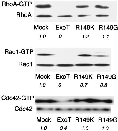
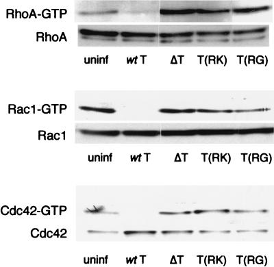
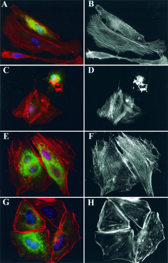
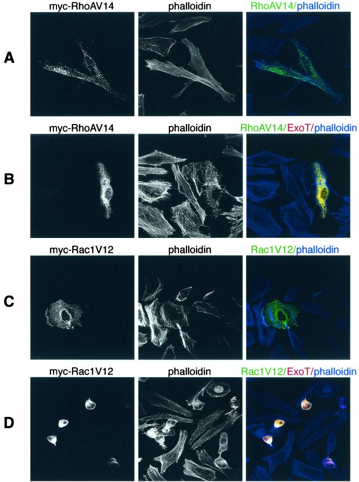
The Pseudomonas aeruginosa protein ExoT is a bacterial GTPase-activating protein (GAP) that has in vitro activity toward Rho, Rac, and Cdc42 GTPases. Expression of ExoT both inhibits the internalization of strain PA103 by macrophages and epithelial cells and is associated with morphological changes (cell rounding and detachment) of infected cells. We find that expression of ExoT leads to the loss of GTP-bound RhoA, Rac1, and Cdc42 in transfected HeLa cells, demonstrating that ExoT has GAP activity in vivo toward all three GTPases. GAP activity is absolutely dependent on the presence of arginine at position 149 but is not affected by whether ExoT is expressed in the absence or presence of other P. aeruginosa type III secreted proteins. We also demonstrate that expression of ExoT in epithelial cells is sufficient to cause stress fiber disassembly by means of ExoT's GAP activity toward RhoA.


Figures

FIG. 1.
FIG. 1.
Cells transfected with wild-type ExoT but not with ExoT(R149G) or ExoT(R149K) show loss of GTP-bound RhoA, Rac1, and Cdc42. HeLa cells were transfected with pMycB-LacZ (Mock), pMycB-T, pMycB-T(R149K), or pMycB-T(R149G) 12 h prior to lysis and determination of total and GTP-bound RhoA, Rac1, and Cdc42 levels as described in the text. (Twenty microliters of lysate was analyzed for total RhoA, Rac1, or Cdc42, while 500 μl of lysate was used in the affinity precipitations.) The experiments shown are characteristic of three or four independent assays. Italicized numbers indicate the fraction of each GTPase present in the GTP-bound form as normalized to mock-transfected cells.
FIG. 2.
FIG. 2.
Infection with ExoT expressing PA103ΔU results in loss of GTP-bound RhoA, Rac1, and Cdc42. HeLa cells were infected with PA103ΔU (MOI = 25 to 50) 0, 1, or 2 h prior to lysis, and determination of total and GTP-bound RhoA, Rac1, and Cdc42 levels was carried out as described in the text. Duplicate samples are shown for each time point. The experiments shown are representative of two or three independent assays.
FIG. 3.
FIG. 3.
Bacterially translocated ExoT(R149G) and ExoT(R149K) do not cause the loss of GTP-bound RhoA, Rac1, or Cdc42 in infected cells. HeLa cells were infected with PA103ΔU (wt T), PA103ΔUΔT (ΔT), PA103ΔU/T(R149K) [T(RK)], or PA103ΔU/T(R149G) [T(RG)] at an MOI of 25 to 50 for 2 h prior to cell lysis. Uninfected cells (uninf) were processed in parallel as a control. Total and GTP-bound RhoA, Rac1, and Cdc42 were detected as described in the text. The experiments shown are representative of two or three independent assays.
FIG.4.
FIG.4.
Cells transfected with wild-type ExoT but not ExoT(R149G) or ExoT(R149K) exhibit stress fiber loss and cell rounding. HeLa cells plated on coverslips were transfected with pMyc-T (A to D), pMyc-T(R149G) (E to F), or pMyc-T(R149K) (G to H) 10 to 14 h prior to fixation. All samples were stained with anti-Myc MAb 9E10 (green) to identify cells expressing ExoT, Texas red-phalloidin (red) to visualize the actin cytoskeleton, and DAPI (blue) to visualize nuclei. Samples were imaged on a Nikon Eclipse E800 microscope equipped with DAPI, fluorescein isothiocyanate, and rhodamine filter sets using a 100× objective, captured with a Spot charge-coupled device camera (Diagnostic Instruments, Inc.) using Spot Advanced 3.0.4 software, pseudocolored, and assembled into figures using Adobe Photoshop 5.0. Panels A, C, E, and G show triply stained cells; panels B, D, F, and H show phalloidin staining only. In panels A to D, cells expressing ExoT show clear loss of stress fibers; cells transfected with either point mutant (E to H) maintain normal stress fiber morphology.
FIG. 5.
FIG. 5.
Expression of RhoAV14 but not of Rac1V12 prevents ExoT-mediated actin cytoskeleton disassembly and cell rounding. HeLa cells plated on coverslips were transfected with 1 μg of either pMyc-RhoAV14 (A and B) or pMyc-Rac1V12 (C and D) for 14 h prior to fixation; 1 μg of pHA-T was cotransfected in panels B and D. All samples were stained with anti-Myc MAb 9E10 (green) and anti-HA MAb 3F10 (red) to identify cells expressing RhoAV14 or Rac1V12 and ExoT, respectively. The actin cytoskeleton was visualized with Bodipy 650/655-conjugated phalloidin (blue). Cells were imaged with a Bio-Rad 1024 confocal microscope equipped with KrAg and HeNe lasers and fluorescein isothiocyanate, Texas red, and Cy5 filter sets using a 60× objective. Z sections were acquired sequentially and processed using NIH Image 1.62b software and were then merged and pseudocolored in Adobe Photoshop 5.0. Cells doubly transfected with ExoT and RhoAV14 (B), which appear yellow, maintained a spread-out shape and showed some stress fibers; cotransfection with Rac1V12, however, did not prevent cell rounding and complete stress fiber loss (yellow-stained cells, panel D).

Similar articles

Cited by

References

    1. Andor, A., K. Trulzsch, M. Essler, A. Roggenkamp, A. Wiedemann, J. Heesemann, and M. Aepfelbacher. 2001. YopE of Yersinia, a GAP for Rho GTPases, selectively modulates Rac-dependent actin structures in endothelial cells. Cell. Microbiol. 3:301-310. - PubMed
    1. Apodaca, G., M. Bomsel, R. Lindstedt, J. Engel, D. Frank, K. E. Mostov, and J. Wiener-Kronish. 1995. Characterization of Pseudomonas aeruginosa-induced MDCK cell injury: glycosylation-defective host cells are resistant to bacterial killing. Infect. Immun. 63:1541-1551. - PMC - PubMed
    1. Bagrodia, S., S. J. Taylor, K. A. Jordon, L. Van Aelst, and R. A. Cerione. 1998. A novel regulator of p21-activated kinases. J. Biol. Chem. 273:23633-23636. - PubMed
    1. Black, D. B., and J. B. Bliska. 2000. The RhoGAP activity of the Yersinia cytotoxin YopE is required for antiphagocytic function and virulence. Mol. Microbiol. 37:515-527. - PubMed
    1. Bliska, J. B. 2000. Yop effectors of Yersinia spp. and actin rearrangements. Trends Microbiol. 8:205-208. - PubMed

Publication types