Review
. 2002 Mar;55(3):162-76.
doi: 10.1136/jcp.55.3.162.

Hodgkin's lymphoma: the pathologist's viewpoint

Affiliations
Review

Hodgkin's lymphoma: the pathologist's viewpoint

S A Pileri et al. J Clin Pathol. 2002 Mar.

Abstract

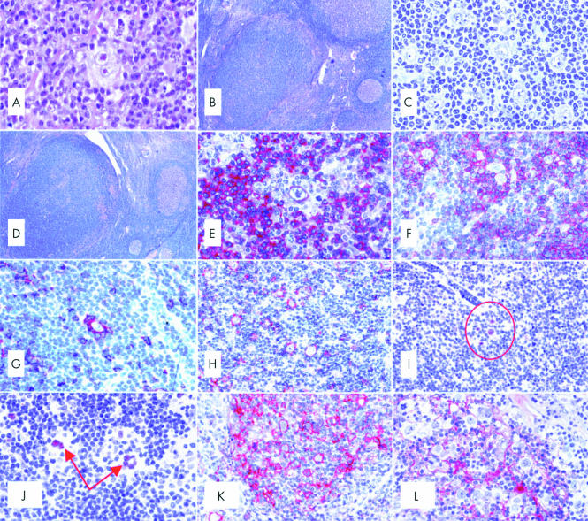
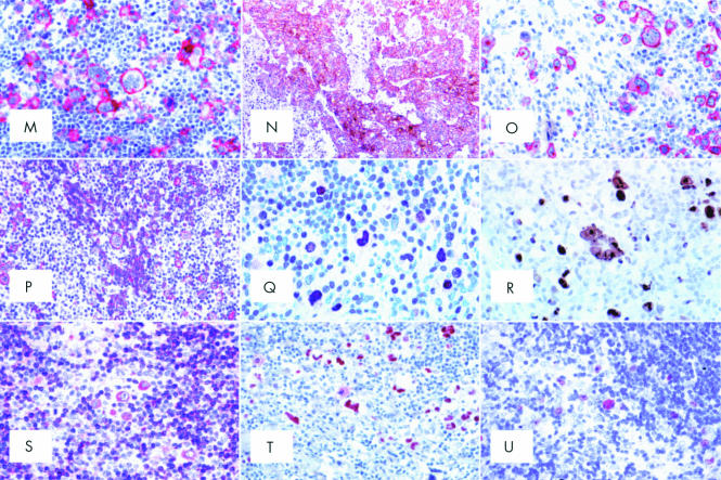
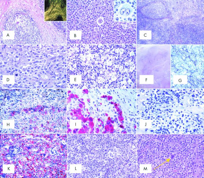
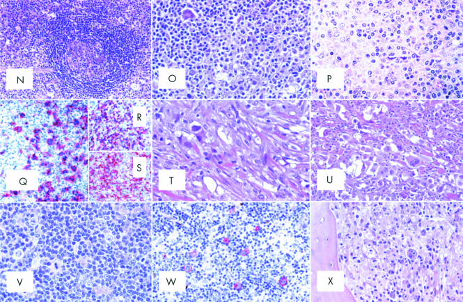
Despite its well known histological and clinical features, Hodgkin's lymphoma (HL) has recently been the object of intense research activity, leading to a better understanding of its phenotype, molecular characteristics, histogenesis, and possible mechanisms of lymphomagenesis. There is complete consensus on the B cell derivation of the tumour in most cases, and on the relevance of Epstein-Barr virus infection and defective cytokinesis in at least a proportion of patients. The REAL/WHO classification recognises a basic distinction between lymphocyte predominance HL (LP-HL) and classic HL (CHL), reflecting the differences in clinical presentation and behaviour, morphology, phenotype, and molecular features. CHL has been classified into four subtypes: lymphocyte rich, nodular sclerosing, with mixed cellularity, and lymphocyte depleted. The borders between CHL and anaplastic large cell lymphoma have become sharper, whereas those between LP-HL and T cell rich B cell lymphoma remain ill defined. Treatments adjusted to the pathobiological characteristics of the tumour in at risk patients have been proposed and are on the way to being applied.


Figures

Figure 1
Figure 1
(A) A typical diagnostic Reed-Sternberg (RS) cell within a composite inflammatory milieu (haematoxylin and eosin; original magnification, ×500). (B) Nodular lymphocyte predominant Hodgkin's lymphoma (LP-HL): at low power, a neoplastic nodule can be seen as a densely packed cellular area with a high content of small lymphocytes (Giemsa; original magnification, ×40). (C) Nodular LP-HL: at higher magnification, some popcorn cells and one Reed-Sternberg-like element are detected among small lymphocytes (Giemsa; original magnification, ×450). (D) Nodular LP-HL: at low magnification, a progressively transformed germinal centre looks like a nodule of LP-HL; however, among small lymphocytes there are some centroblasts and centrocytes, but no popcorn cells (Giemsa; original magnification, ×40). (E) Nodular LP-HL: a popcorn cell and most small lymphocytes within a nodule express the B cell marker CD79a (APAAP technique, Gill's haematoxylin counterstain; original magnification, ×500). (F) Nodular LP-HL: popcorn cells are surrounded by rosettes of CD3+ T cells (APAAP technique, Gill's haematoxylin counterstain; original magnification, ×250). (G) Nodular LP-HL: rosetting T cells largely express CD57 (APAAP technique, Gill's haematoxylin counterstain; original magnification, ×250). (H) Nodular LP-HL: popcorn cells express epithelial membrane antigen (EMA) (APAAP technique, Gill's haematoxylin counterstain; original magnification, ×250). (I) Nodular LP-HL: popcorn cells (circled) express the bcl-6 gene product (APAAP technique, Gill's haematoxylin counterstain; original magnification, ×250). (J) Nodular LP-HL: positivity of neoplastic elements for the Oct2 gene product (APAAP technique, Gill's haematoxylin counterstain; original magnification, ×300). (K) Nodular LP-HL: popcorn cells strongly express CD40 (APAAP technique, Gill's haematoxylin counterstain; original magnification, ×300). (L) Nodular LP-HL: neoplastic elements are found within a delicate meshwork of CD21+ follicular dendritic cells (APAAP technique, Gill's haematoxylin counterstain; original magnification, ×300). (M) Common Hodgkin's lymphoma (CHL): Hodgkin and Reed-Sternberg (H&RS) cells express the CD30 molecule both at the cytoplasmic membrane and in the Golgi area (dot-like positivity) (APAAP technique, Gill's haematoxylin counterstain; original magnification, ×350). (N) CD30 membrane bound positivity in an example of embryonal carcinoma (immunoperoxidase, SABC technique; Gill's haematoxylin counterstain; original magnification, ×200). (O) CHL: neoplastic cells show membrane bound and dot-like CD15 positivity (APAAP technique, Gill's haematoxylin counterstain; original magnification, ×350). (P) CHL: H&RS cells display variable degrees of CD20 staining (APAAP technique, Gill's haematoxylin counterstain; original magnification, ×200). (Q) CHL: staining for IRF4/MUM1 gene product (APAAP technique, Gill's haematoxylin counterstain; original magnification, ×350). (R) CHL: most neoplastic cells express the Ki-67 molecule as revealed by the Mib-1 monoclonal antibody; some small lymphocytes are also in the cell cycle (immunoperoxidase, SABC technique; Gill's haematoxylin counterstain; original magnification, ×500). (S) CHL: moderate Bcl-2 protein expression by H&RS cells and a ratio of small lymphocytes (APAAP technique, Gill's haematoxylin counterstain; original magnification, ×300). (T) CHL: most neoplastic cells show p53 overexpression (APAAP technique, Gill's haematoxylin counterstain; original magnification, ×300). (U) CHL: H & RS show genomic Epstein-Barr virus integration by in situ hybridisation with fluorescein labelled EBER1/2 probes (APAAP technique, Gill's haematoxylin counterstain; original magnification, ×300).
Figure 1
Figure 1
(A) A typical diagnostic Reed-Sternberg (RS) cell within a composite inflammatory milieu (haematoxylin and eosin; original magnification, ×500). (B) Nodular lymphocyte predominant Hodgkin's lymphoma (LP-HL): at low power, a neoplastic nodule can be seen as a densely packed cellular area with a high content of small lymphocytes (Giemsa; original magnification, ×40). (C) Nodular LP-HL: at higher magnification, some popcorn cells and one Reed-Sternberg-like element are detected among small lymphocytes (Giemsa; original magnification, ×450). (D) Nodular LP-HL: at low magnification, a progressively transformed germinal centre looks like a nodule of LP-HL; however, among small lymphocytes there are some centroblasts and centrocytes, but no popcorn cells (Giemsa; original magnification, ×40). (E) Nodular LP-HL: a popcorn cell and most small lymphocytes within a nodule express the B cell marker CD79a (APAAP technique, Gill's haematoxylin counterstain; original magnification, ×500). (F) Nodular LP-HL: popcorn cells are surrounded by rosettes of CD3+ T cells (APAAP technique, Gill's haematoxylin counterstain; original magnification, ×250). (G) Nodular LP-HL: rosetting T cells largely express CD57 (APAAP technique, Gill's haematoxylin counterstain; original magnification, ×250). (H) Nodular LP-HL: popcorn cells express epithelial membrane antigen (EMA) (APAAP technique, Gill's haematoxylin counterstain; original magnification, ×250). (I) Nodular LP-HL: popcorn cells (circled) express the bcl-6 gene product (APAAP technique, Gill's haematoxylin counterstain; original magnification, ×250). (J) Nodular LP-HL: positivity of neoplastic elements for the Oct2 gene product (APAAP technique, Gill's haematoxylin counterstain; original magnification, ×300). (K) Nodular LP-HL: popcorn cells strongly express CD40 (APAAP technique, Gill's haematoxylin counterstain; original magnification, ×300). (L) Nodular LP-HL: neoplastic elements are found within a delicate meshwork of CD21+ follicular dendritic cells (APAAP technique, Gill's haematoxylin counterstain; original magnification, ×300). (M) Common Hodgkin's lymphoma (CHL): Hodgkin and Reed-Sternberg (H&RS) cells express the CD30 molecule both at the cytoplasmic membrane and in the Golgi area (dot-like positivity) (APAAP technique, Gill's haematoxylin counterstain; original magnification, ×350). (N) CD30 membrane bound positivity in an example of embryonal carcinoma (immunoperoxidase, SABC technique; Gill's haematoxylin counterstain; original magnification, ×200). (O) CHL: neoplastic cells show membrane bound and dot-like CD15 positivity (APAAP technique, Gill's haematoxylin counterstain; original magnification, ×350). (P) CHL: H&RS cells display variable degrees of CD20 staining (APAAP technique, Gill's haematoxylin counterstain; original magnification, ×200). (Q) CHL: staining for IRF4/MUM1 gene product (APAAP technique, Gill's haematoxylin counterstain; original magnification, ×350). (R) CHL: most neoplastic cells express the Ki-67 molecule as revealed by the Mib-1 monoclonal antibody; some small lymphocytes are also in the cell cycle (immunoperoxidase, SABC technique; Gill's haematoxylin counterstain; original magnification, ×500). (S) CHL: moderate Bcl-2 protein expression by H&RS cells and a ratio of small lymphocytes (APAAP technique, Gill's haematoxylin counterstain; original magnification, ×300). (T) CHL: most neoplastic cells show p53 overexpression (APAAP technique, Gill's haematoxylin counterstain; original magnification, ×300). (U) CHL: H & RS show genomic Epstein-Barr virus integration by in situ hybridisation with fluorescein labelled EBER1/2 probes (APAAP technique, Gill's haematoxylin counterstain; original magnification, ×300).
Figure 2
Figure 2
(A) Nodular sclerosing common Hodgkin's lymphoma (NS-CHL): the normal lymph node structure is largely effaced because of a nodular growth; the nodule is surrounded by thick collagen bands originating from the capsule (haematoxylin and eosin; original magnification, ×40). Inset: birefringence of collagen bands (polarised light microscopy; original magnification, ×20). (B) NS-CHL: scattered lacunar cells can easily be seen within one nodule (haematoxylin and eosin; original magnification, ×300). Inset: cytological details of a lacunar cell (Giemsa; original magnification, ×800). (C) NS-CHL: so called cellular phase; note the nodularity of the growth (haematoxylin and eosin; original magnification, ×80). (D) NS-CHL: so called syncytial variant; note the cohesive growth pattern of neoplastic cells (haematoxylin and eosin; original magnification, ×400). (E) NS-CHL: an example of grade II tumour; note the content of neoplastic cells within the nodule, which covers more than 25% of the examined area (haematoxylin and eosin; original magnification, ×300). (F) An example of anaplastic large cell lymphoma of the Hodgkin-like type (ALCL-HL): the tumour consists of nodules partly surrounded by collagen bands (haematoxylin and eosin, original magnification, ×20).(G) ALCL-HL: at higher magnification it can be seen that the collagen bands are almost exclusively formed by neoplastic cells (Giemsa; original magnification, ×300). (H) ALCL-HL: typical intrasinusoidal diffusion of neoplastic cells as shown by CD30 staining (APAAP technique, Gill's haematoxylin counterstain; original magnification, ×200). (I) ALCL-HL: ALK protein expression by neoplastic cells (APAAP technique, Gill's haematoxylin counterstain; original magnification, ×400). (J) Primary mediastinal large B cell lymphoma (PMLBCL): neoplastic cells sometimes have multiple nuclei, show a wide rim of clear, fragile cytoplasm, and elicit a stromal reaction with compartmentalisation (Giemsa; original magnification, ×300). (K) PMLBCL: neoplastic cells express the CD30 molecule (APAAP technique, Gill's haematoxylin counterstain; original magnification, ×300). (L) Mixed cellularity common Hodgkin's lymphoma (MC-CHL): Hodgkin and Reed-Sternberg (H&RS) cells are easily identified; they are found with a cellular milieu consisting of small lymphocytes, some plasma cells, histiocytes, and granulocytes (haematoxylin and eosin; original magnification, ×350). (M) MC-CHL: an example of a mummified cell (arrow) (haematoxylin and eosin; original magnification, ×350). (N) MC-CHL: the tumour has a patent interfollicular location; a spared follicle with Castleman-like features can be seen (haematoxylin and eosin; original magnification, ×150). (O) MC-CHL: the tumour contains reactive epithelioid cells (haematoxylin and eosin; original magnification, ×300). (P) Peripheral T cell lymphoma not otherwise specified with a high content of epithelioid cells (so called Lennert's lymphoma): lymphoid elements show variation in size and shape (haematoxylin and eosin; original magnification, ×400). (Q) T cell rich, histiocyte rich large B cell lymphoma (TCRBCL): neoplastic cells strongly express the CD79a molecule (APAAP technique, Gill's haematoxylin counterstain; original magnification, ×300). (R) TCRBCL: reactive T cells and histiocytes are stained by the anti-CD3 antibody (APAAP technique, Gill's haematoxylin counterstain; original magnification, ×150). (S) TCRBCL: reactive T cells and histiocytes are stained by the anti-CD68 antibody (APAAP technique, Gill's haematoxylin counterstain; original magnification, ×150). (T) Lymphocyte depleted common Hodgkin's lymphoma (LD-CHL), fibrotic variant: rare neoplastic cells are surrounded by thick collagen bands with a haphazard organisation, some histiocytes and scanty lymphocytes (haematoxylin and eosin; original magnification, ×400). (U) LD-CHL, sarcomatous variant: H&RS cells are quite numerous; there is a certain degree of fibrotic reaction; small lymphocytes are exceedingly rare (haematoxylin and eosin; original magnification, ×350). (V) Lymphocyte rich common Hodgkin's lymphoma (LR-CHL): mononuclear and diagnostic neoplastic elements are found within a cellular milieu mostly consisting of small lymphocytes (haematoxylin and eosin; original magnification, ×350). (W) LR-CHL: neoplastic cells express CD15 (APAAP technique, Gill's haematoxylin counterstain; original magnification, ×300). (X) CHL: typical example of bone marrow involvement; note the fibrotic reaction, and the presence of H&RS cells (haematoxylin and eosin; original magnification, ×300).
Figure 2
Figure 2
(A) Nodular sclerosing common Hodgkin's lymphoma (NS-CHL): the normal lymph node structure is largely effaced because of a nodular growth; the nodule is surrounded by thick collagen bands originating from the capsule (haematoxylin and eosin; original magnification, ×40). Inset: birefringence of collagen bands (polarised light microscopy; original magnification, ×20). (B) NS-CHL: scattered lacunar cells can easily be seen within one nodule (haematoxylin and eosin; original magnification, ×300). Inset: cytological details of a lacunar cell (Giemsa; original magnification, ×800). (C) NS-CHL: so called cellular phase; note the nodularity of the growth (haematoxylin and eosin; original magnification, ×80). (D) NS-CHL: so called syncytial variant; note the cohesive growth pattern of neoplastic cells (haematoxylin and eosin; original magnification, ×400). (E) NS-CHL: an example of grade II tumour; note the content of neoplastic cells within the nodule, which covers more than 25% of the examined area (haematoxylin and eosin; original magnification, ×300). (F) An example of anaplastic large cell lymphoma of the Hodgkin-like type (ALCL-HL): the tumour consists of nodules partly surrounded by collagen bands (haematoxylin and eosin, original magnification, ×20).(G) ALCL-HL: at higher magnification it can be seen that the collagen bands are almost exclusively formed by neoplastic cells (Giemsa; original magnification, ×300). (H) ALCL-HL: typical intrasinusoidal diffusion of neoplastic cells as shown by CD30 staining (APAAP technique, Gill's haematoxylin counterstain; original magnification, ×200). (I) ALCL-HL: ALK protein expression by neoplastic cells (APAAP technique, Gill's haematoxylin counterstain; original magnification, ×400). (J) Primary mediastinal large B cell lymphoma (PMLBCL): neoplastic cells sometimes have multiple nuclei, show a wide rim of clear, fragile cytoplasm, and elicit a stromal reaction with compartmentalisation (Giemsa; original magnification, ×300). (K) PMLBCL: neoplastic cells express the CD30 molecule (APAAP technique, Gill's haematoxylin counterstain; original magnification, ×300). (L) Mixed cellularity common Hodgkin's lymphoma (MC-CHL): Hodgkin and Reed-Sternberg (H&RS) cells are easily identified; they are found with a cellular milieu consisting of small lymphocytes, some plasma cells, histiocytes, and granulocytes (haematoxylin and eosin; original magnification, ×350). (M) MC-CHL: an example of a mummified cell (arrow) (haematoxylin and eosin; original magnification, ×350). (N) MC-CHL: the tumour has a patent interfollicular location; a spared follicle with Castleman-like features can be seen (haematoxylin and eosin; original magnification, ×150). (O) MC-CHL: the tumour contains reactive epithelioid cells (haematoxylin and eosin; original magnification, ×300). (P) Peripheral T cell lymphoma not otherwise specified with a high content of epithelioid cells (so called Lennert's lymphoma): lymphoid elements show variation in size and shape (haematoxylin and eosin; original magnification, ×400). (Q) T cell rich, histiocyte rich large B cell lymphoma (TCRBCL): neoplastic cells strongly express the CD79a molecule (APAAP technique, Gill's haematoxylin counterstain; original magnification, ×300). (R) TCRBCL: reactive T cells and histiocytes are stained by the anti-CD3 antibody (APAAP technique, Gill's haematoxylin counterstain; original magnification, ×150). (S) TCRBCL: reactive T cells and histiocytes are stained by the anti-CD68 antibody (APAAP technique, Gill's haematoxylin counterstain; original magnification, ×150). (T) Lymphocyte depleted common Hodgkin's lymphoma (LD-CHL), fibrotic variant: rare neoplastic cells are surrounded by thick collagen bands with a haphazard organisation, some histiocytes and scanty lymphocytes (haematoxylin and eosin; original magnification, ×400). (U) LD-CHL, sarcomatous variant: H&RS cells are quite numerous; there is a certain degree of fibrotic reaction; small lymphocytes are exceedingly rare (haematoxylin and eosin; original magnification, ×350). (V) Lymphocyte rich common Hodgkin's lymphoma (LR-CHL): mononuclear and diagnostic neoplastic elements are found within a cellular milieu mostly consisting of small lymphocytes (haematoxylin and eosin; original magnification, ×350). (W) LR-CHL: neoplastic cells express CD15 (APAAP technique, Gill's haematoxylin counterstain; original magnification, ×300). (X) CHL: typical example of bone marrow involvement; note the fibrotic reaction, and the presence of H&RS cells (haematoxylin and eosin; original magnification, ×300).

References

    1. Hummel M, Marafioti T, Stein H. Immunoglobulin V genes in Reed-Sternberg cells. N Engl J Med 1996;334:405–6. - PubMed
    1. Marafioti T, Hummel M, Anagnostopoulos I, et al. Origin of nodular lymphocyte predominant Hodgkin's disease from a clonal expansion of highly mutated germinal center B cells. N Engl J Med 1997;337:453–8. - PubMed
    1. Izban KF, Nawrocki JF, Alkan S, et al. Monoclonal IgH gene rearrangement in microdissected nodules from nodular sclerosis Hodgkin's disease. Am J Clin Pathol 1998;110:599–606. - PubMed
    1. Braeuninger A, Hansmann M-L, Strickler JG, et al. Identification of common germinal-center B-cell precursors in two patients with both Hodgkin's disease and non-Hodgkin's lymphoma. N Engl J Med 1999;340:1239–47. - PubMed
    1. Küppers R, Klein U, Hansmann M-L, et al. Cellular origin of human B-cell lymphomas. N Engl J Med 1999;341:1520–9. - PubMed

Publication types