

Intermediate grade osteosarcoma and chondrosarcoma arising in an osteochondroma. A case report of a patient with hereditary multiple exostoses
- PMID: 11896078
- PMCID: PMC1769605
- DOI: 10.1136/jcp.55.3.226
Intermediate grade osteosarcoma and chondrosarcoma arising in an osteochondroma. A case report of a patient with hereditary multiple exostoses
Abstract
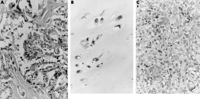
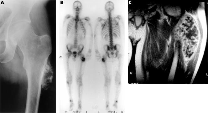
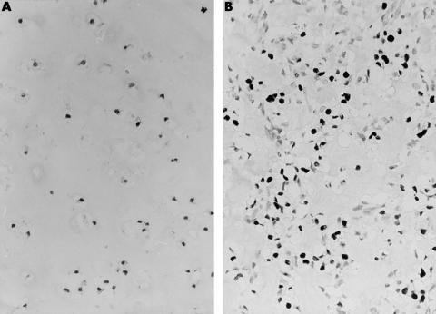
A 40 year old man with hereditary multiple exostoses (HME), affecting predominantly his left proximal tibia, distal femur, and proximal femur, underwent resection of an osteochondroma near the trochanter major of his left proximal femur because of malignant transformation of the cartilaginous cap towards secondary peripheral chondrosarcoma. The patient had a history of a papillary thyroid carcinoma four years previously. At examination of the resected specimen, a third malignant tumour, an intermediate grade osteosarcoma (grade II/IV), was found in the osseous stalk of the osteochondroma. Although no mutations were found in the EXT1 and EXT2 genes, the genes involved in HME, or in exons 5-8 of the p53 gene, the development of three malignancies before the age of 40 suggests that this patient is genetically prone to malignant transformation.
Figures
References
-
- Ahn J, Ludecke H-J, Lindow S, et al. Cloning of the putative tumour suppressor gene for hereditary multiple exostoses (EXT1). Nat Genet 1995;11:137–43. - PubMed
-
- Stickens D, Clines G, Burbee D, et al. The EXT2 multiple exostoses gene defines a family of putative tumour suppressor genes. Nat Genet 1996;14:25–32. - PubMed
-
- Wuyts W, Van Hul W, Wauters J, et al. Positional cloning of a gene involved in hereditary multiple exostoses. Hum Mol Genet 1996;5:1547–57. - PubMed
-
- Devilee P, Van den Broek M, Kuipers-Dijkshoorn N, et al. At least four different chromosomal regions are involved in loss of heterozygosity in human breast carcinoma. Genomics 1989;5:554–60. - PubMed
Publication types
MeSH terms
Substances
LinkOut - more resources
Full Text Sources
Medical
Research Materials
Miscellaneous