. 2002 Mar 15;22(6):2054-62.
doi: 10.1523/JNEUROSCI.22-06-02054.2002.

Long-term depression in the adult hippocampus in vivo involves activation of extracellular signal-regulated kinase and phosphorylation of Elk-1

Affiliations

Long-term depression in the adult hippocampus in vivo involves activation of extracellular signal-regulated kinase and phosphorylation of Elk-1

Edda Thiels et al. J Neurosci. .

Abstract

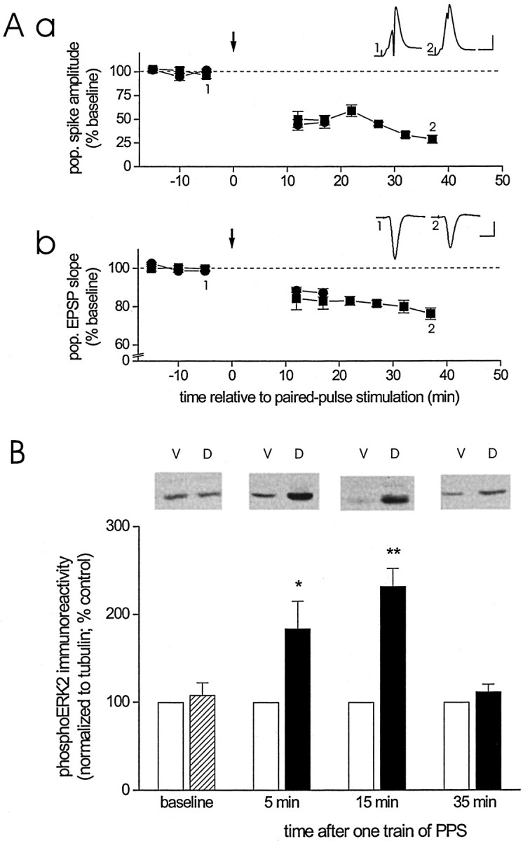
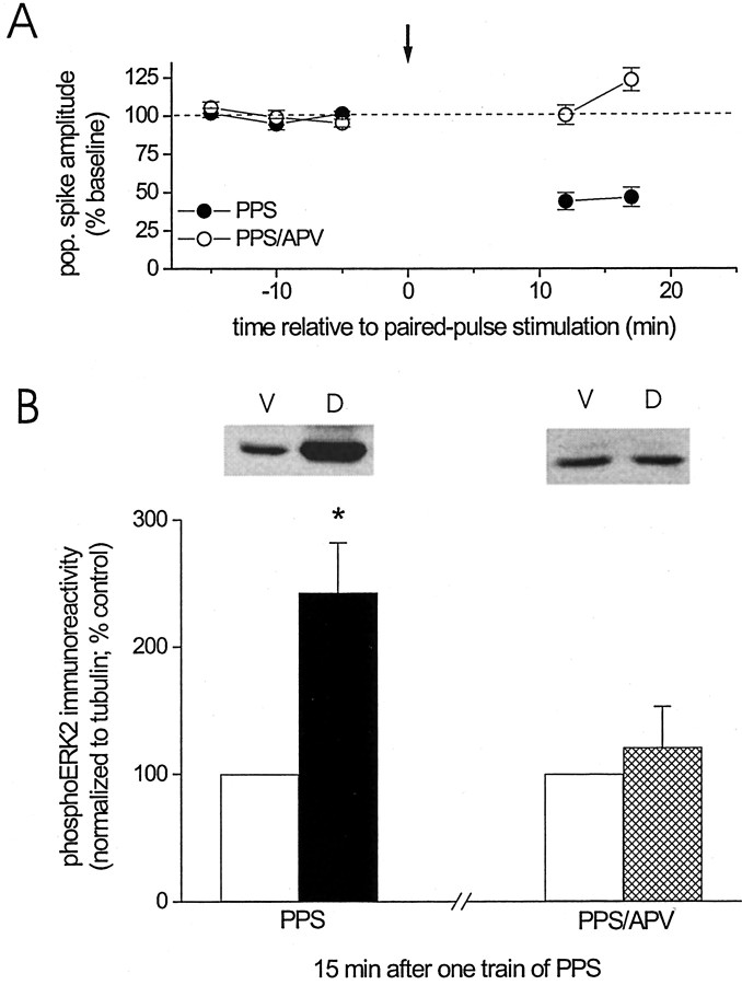
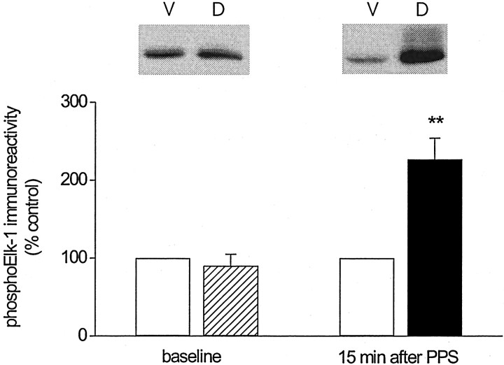
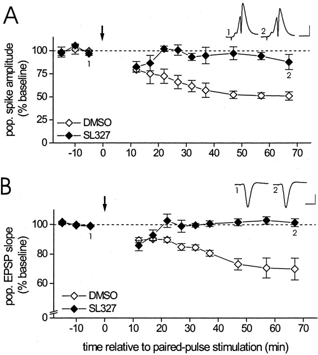
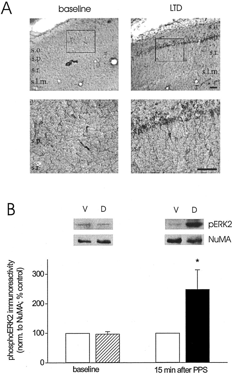
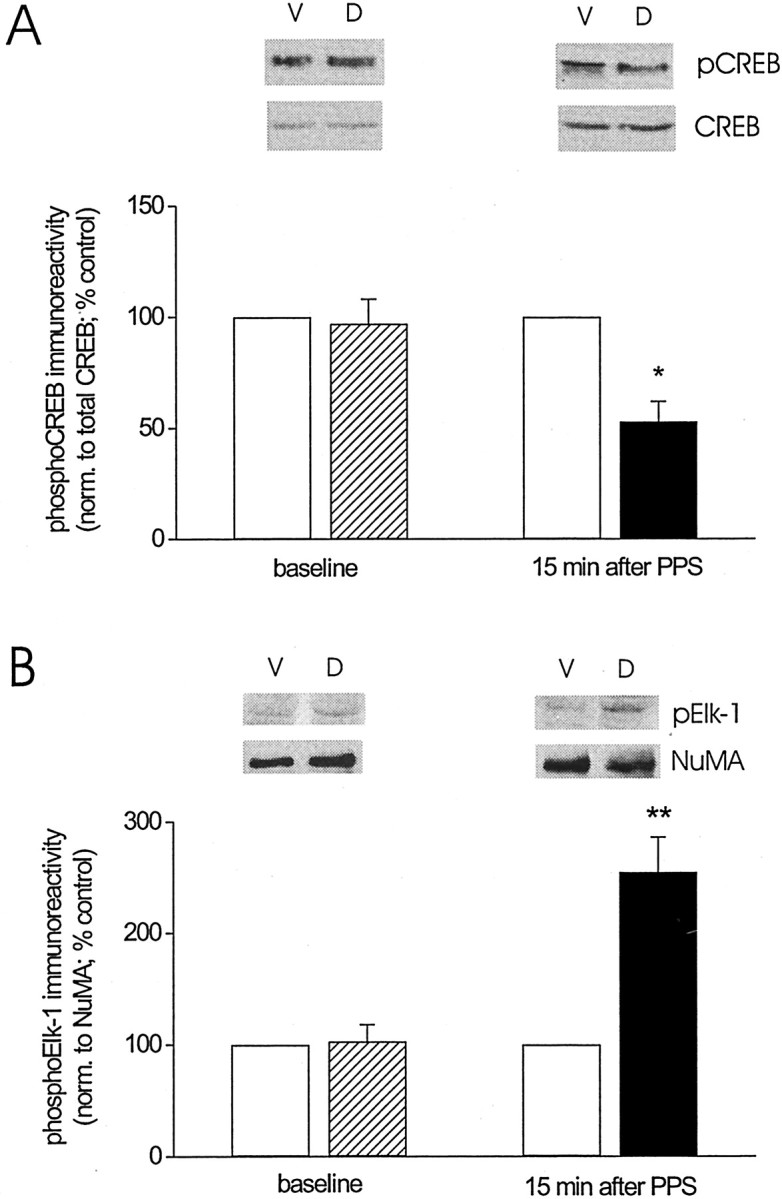
Protein kinase cascades likely play a critical role in the signaling events that underlie synaptic plasticity and memory. The extracellular signal-regulated kinase (ERK) cascade is suited well for such a role because its targets include regulators of gene expression. Here we report that the ERK cascade is recruited during long-term depression (LTD) of synaptic strength in area CA1 of the adult hippocampus in vivo and selectively impacts on phosphorylation of the nuclear transcription factor Elk-1. Using a combination of in vivo electrophysiology, biochemistry, pharmacology, and immunohistochemistry, we found the following: (1) ERK phosphorylation, including phosphorylation of nuclear ERK, and ERK phosphotransferase activity are increased markedly, albeit transiently, after the induction of NMDA receptor-dependent LTD at the commissural input to area CA1 pyramidal cells in the hippocampus of anesthetized adult rats; (2) LTD-inducing paired-pulse stimulation fails to produce lasting LTD in the presence of the ERK kinase inhibitor SL327, which suggests that ERK activation is necessary for the persistence of LTD; and (3) ERK activation during LTD results in increased phosphorylation of Elk-1 but not of the transcription factor cAMP response element-binding protein. Our findings indicate that the ERK cascade transduces signals from the synapse to the nucleus during LTD in hippocampal area CA1 in vivo, as it does during long-term potentiation in area CA1, but that the pattern of coupling of the ERK cascade to transcriptional regulators differs between the two forms of synaptic plasticity.


Figures

Fig. 1.
Fig. 1.
LTD in area CA1 of the adult hippocampusin vivo is associated with an increase in activated ERK2. Aa, Group data (mean ± SEM) of the amplitude of the CA1 pyramidal cell population spike evoked by stimulation of commissural fibers before and after delivery of one train of paired pulses (downward arrow) to these fibers. The data are expressed as a percentage of the average population spike amplitude before paired-pulse stimulation. Animals were killed either 15 min (circles; n = 6) or 35 min (squares; n = 5) after termination of paired-pulse stimulation. Inset, Average of 10 waveforms of population spikes recorded in the same animal before (1) and after (2) paired-pulse stimulation at the times indicated. Calibration: 2 mV, 10 msec. Ab, Similar group data of the initial slope of the pyramidal cell population EPSP recorded in stratum radiatum before and after paired-pulse stimulation (downward arrow). Animals were killed at the same times after termination of the stimulation train as described above (15 min, circles,n = 6; 35 min, squares,n = 5). Inset, Averaged waveforms of population EPSPs recorded before and after paired-pulse stimulation, as described above (calibration as above). B, Group data (mean ± SEM) of dual-phosphorylated ERK2 immunoreactivity, normalized to tubulin, for ventral area CA1 homogenates (control,open bars) and dorsal area CA1 homogenates (baseline,striped bar; LTD, filled bars) derived from animals killed either 5 min after termination of baseline recording (baseline; n = 12) or 5 (n = 8), 15 (n = 12), or 35 (n = 10) min after termination of PPS. Data are expressed as a percentage of dual-phosphorylated ERK2 immunoreactivity detected in ventral area CA1 homogenates. Dual-phosphorylated ERK2 immunoreactivity was normalized to tubulin immunoreactivity rather than total ERK immunoreactivity, because the capability of nonphospho-specific ERK antibodies to bind to ERK was found to be reduced after the induction of LTD (Norman et al., 2000). Evidence indicates that total ERK level is unaffected by the induction of LTD (Norman et al., 2000). Representative Western blots of dual-phosphorylated ERK2 for ventral (V) and dorsal (D) area CA1 homogenate for the time points indicated on the x-axis. Asterisksindicate significant difference between control and LTD samples at the indicated time points (Student's t test for matched samples; *p < 0.05; **p < 0.01).
Fig. 2.
Fig. 2.
Blockade of NMDA receptors prevents the induction of LTD, as well as the associated increase in activated ERK2.A, Group data (mean ± SEM) of the amplitude of the population spike recorded before and after paired-pulse stimulation (downward arrow) during continuous administration of the specific NMDA receptor antagonist d-APV (100 μm in the drug pipette; open circles;n = 5). For purposes of comparison, the effect of paired-pulse stimulation in the absence of d-APV is depicted as well (filled circles;n = 6; data are taken from Fig.1Aa). B, Group data (mean ± SEM) of dual-phosphorylated ERK2 immunoreactivity, normalized to tubulin, for ventral area CA1 homogenates (control, open bars) and dorsal area CA1 homogenates (filled or cross-hatched bar) derived from animals who received paired-pulse stimulation in either the presence of d-APV (cross-hatched bar;n = 5) or the absence of the drug (filled bar; n = 6) and were killed 15 min after termination of PPS. Data are expressed as a percentage of dual-phosphorylated ERK2 immunoreactivity detected in ventral area CA1 homogenates. Representative Western blots of dual-phosphorylated ERK2 for ventral (V) and dorsal (D) area CA1 homogenate for the two conditions indicated on the x-axis.Asterisk indicates significant difference between ventral and dorsal samples (Student's t test for matched samples; *p < 0.05).
Fig. 3.
Fig. 3.
LTD in area CA1 of the adult hippocampus in vivo is associated with an increase in ERK phosphotransferase activity. Group data (mean ± SEM) of Ser383-phosphorylated Elk-1 immunoreactivity in samples containing exogenously added Elk-1 and dual-phosphorylated ERK immunoprecipitated from dorsal area CA1 homogenate (baseline, striped bar; LTD, filled bar) and ventral CA1 homogenate (open bars) of animals killed either 5 min after baseline stimulation (baseline; n = 6) or 15 min after termination of paired-pulse stimulation (15 min after PPS; n = 6). Data are expressed as a percentage of Ser383-phosphorylated Elk-1 immunoreactivity detected in samples containing dual-phosphorylated ERK immunoprecipitated from ventral area CA1 homogenates. For additional details, see Results. Representative Western blots of Ser383-phosphorylated Elk-1 for ventral (V) and dorsal (D) area CA1 homogenate for the two time points indicated on thex-axis. Asterisks indicate significant difference between control and LTD samples (Student's ttest for matched samples; **p < 0.01).
Fig. 4.
Fig. 4.
Inhibition of the ERK kinase MEK prevents the expression of LTD in area CA1 of the adult hippocampus in vivo. A, Group data (mean ± SEM) of the amplitude of the CA1 pyramidal cell population spike evoked by stimulation of commissural fibers before and after delivery of one train of paired-pulse stimulation (downward arrow) after animals were treated with either the MEK inhibitor SL327 (50 mg/kg, i.p.; filled diamonds; n = 4) or vehicle solution (100% DMSO, 1 ml/kg, i.p.; open diamonds; n = 4) 1–2 hr before paired-pulse stimulation. For additional details, see Results.Inset, Average of 10 waveforms of population spikes recorded in the same SL327-treated animal before (1) and after (2) paired-pulse stimulation at the times indicated. Calibration: 2 mV, 10 msec. B, Similar group data of the initial slope of the pyramidal cell population EPSP recorded in stratum radiatum before and after paired-pulse stimulation (downward arrow) after treatment with either SL327 (filled diamonds;n = 5) or DMSO (open diamonds;n = 5). Inset, Averaged waveforms of population EPSPs recorded in a SL327-treated animal before and after paired-pulse stimulation, as described above (calibration as above).
Fig. 5.
Fig. 5.
LTD in area CA1 of the adult hippocampus in vivo is associated with an increase in activated ERK in CA1 pyramidal cell dendrites and somata, including nuclei.A, Dual-phosphorylated ERK1/2 staining in sections of area CA1 from an animal that received baseline stimulation only (baseline) and an animal that received baseline and paired-pulse stimulation (LTD). Tissue sections from the two animals were processed simultaneously. Bottom panelsshow the boxed section marked in the respectivetop panels at a higher magnification. Scale bar, 100 μm. Similar patterns of staining and differences in staining intensity after baseline stimulation versus LTD-inducing stimulation were observed in other pairs of animals (n = 4).B, Group data (mean ± SEM) of dual-phosphorylated ERK2 immunoreactivity, normalized to NuMA, for nuclear extracts prepared from ventral area CA1 (control, open bars) and dorsal area CA1 (baseline, striped bar; LTD,filled bar) collected from animals killed either 5 min after termination of baseline recording (baseline;n = 4) or 15 min after termination of paired-pulse stimulation (15 min after PPS; n = 5). Data are expressed as a percentage of dual-phosphorylated ERK2 immunoreactivity detected in nuclear extracts from ventral area CA1. Representative Western blots of dual-phosphorylated ERK2 (pERK2) and of NuMA for nuclear extracts from ventral (V) and dorsal (D) area CA1 for the two time points indicated on thex-axis. Asterisk indicates significant difference between control and LTD samples (Student's ttest for matched samples; *p < 0.05).
Fig. 6.
Fig. 6.
LTD in area CA1 of the adult hippocampus in vivo is associated with a decrease in nuclear CREB phosphorylation but an increase in nuclear Elk-1 phosphorylation.A, Group data (mean ± SEM) of Ser133-phosphorylated CREB immunoreactivity, normalized to total CREB, for nuclear extracts prepared from ventral area CA1 (control,open bars) and dorsal area CA1 (baseline, striped bar; LTD, filled bars) collected from animals killed either 5 min after termination of baseline recording (baseline;n = 5) or 15 min after termination of paired-pulse stimulation (15 min after PPS; n = 6). Data are expressed as a percentage of Ser133-phosphorylated CREB immunoreactivity detected in nuclear extracts from ventral area CA1. Representative Western blots of Ser133-phosphorylated CREB (pCREB) and total CREB for nuclear extracts from ventral (V) and dorsal (D) area CA1 for the two time points indicated on the x-axis (Student's t test for matched samples; *p < 0.05). B, Similar group data for Ser383-phosphorylated Elk-1 immunoreactivity, normalized to NuMA, for nuclear extracts prepared from ventral area CA1 (control,open bars) and dorsal area CA1 (baseline, striped bar; LTD, filled bars) collected from animals killed either 5 min after termination of baseline recording (baseline;n = 4) or 15 min after termination of paired-pulse stimulation (15 min after PPS; n = 7). Representative Western blots of Ser383-phosphorylated Elk-1 (pElk-1) and NuMA for nuclear extracts from ventral (V) and dorsal (D) area CA1 for the two time points indicated on the x-axis. PhosphoElk-1 immunoreactivity was normalized to NuMA immunoreactivity because of an inconsistent detectability of the total Elk-1 signal. In those cases in which total Elk-1 could be detected, no difference was noted between normalization to Elk-1 and to NuMA. Asterisks indicate significant difference between control and LTD samples (Student's t test for matched samples; **p < 0.01).

Similar articles

Cited by

References

    1. Abraham WC, Demmer J, Richardson LC, Williams JM, Lawlor PA, Mason SE, Tate WP, Dragunow M. Correlations between early gene induction and the persistence of LTP. Neuroscience. 1993;56:717–727. - PubMed
    1. Alberini CM, Ghirardi M, Huang Y-Y, Nguyen PV, Kandel ER. A molecular switch for the consolidation of long-term memory: cAMP-inducible gene expression. Ann NY Acad Sci. 1995;758:261–286. - PubMed
    1. Atkins CM, Selcher JC, Petraitis JJ, Trzaskos JM, Sweatt JD. The MAPK cascade is required for mammalian associative learning. Nat Neurosci. 1998;1:602–609. - PubMed
    1. Barria A, Muller D, Derkach V, Griffith LC, Soderling TR. Regulatory phosphorylation of AMPA-type glutamate receptors by CaM-KII during long-term potentiation. Science. 1997;276:2042–2045. - PubMed
    1. Bear MF, Malenka RC. Synaptic plasticity: LTP and LTD. Curr Opin Neurobiol. 1994;4:389–399. - PubMed

Publication types

MeSH terms

Substances

LinkOut - more resources