. 2002 Mar 15;22(6):2125-34.
doi: 10.1523/JNEUROSCI.22-06-02125.2002.

Localization and regulation of the tissue plasminogen activator-plasmin system in the hippocampus

Affiliations

Localization and regulation of the tissue plasminogen activator-plasmin system in the hippocampus

Fernando J Salles et al. J Neurosci. .

Abstract

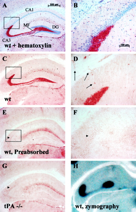
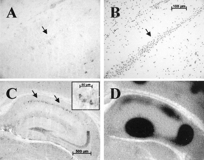
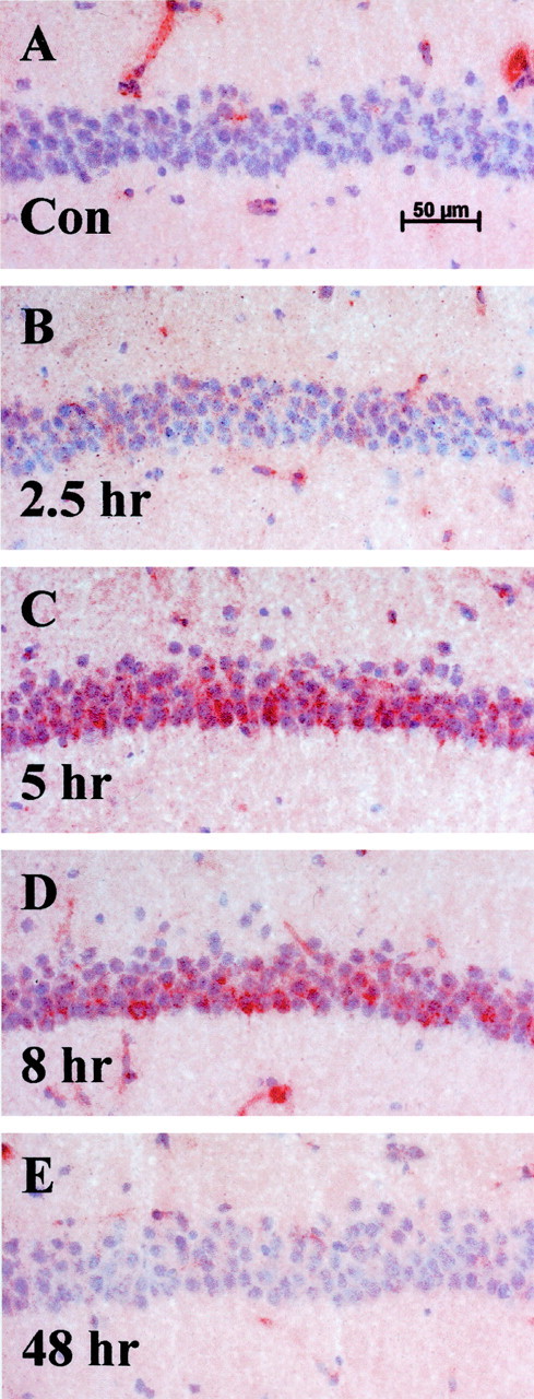
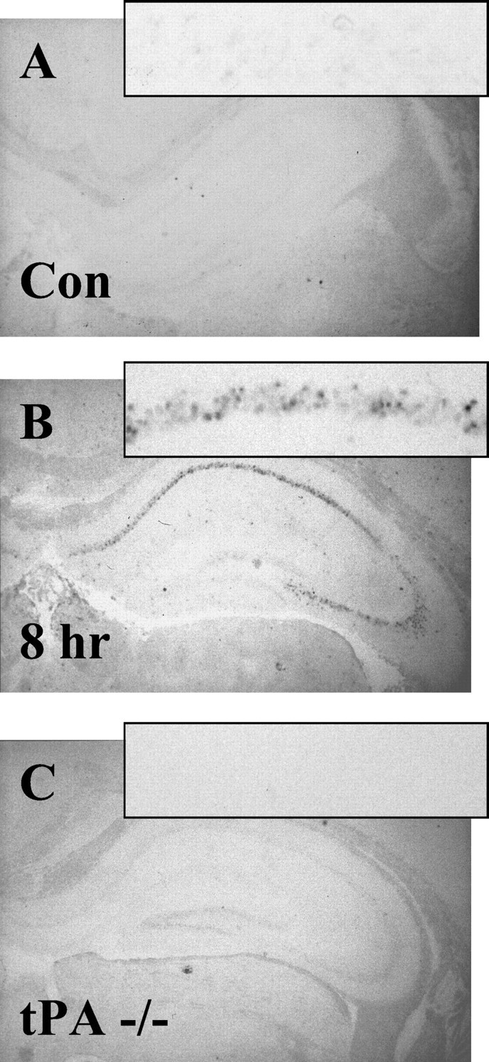
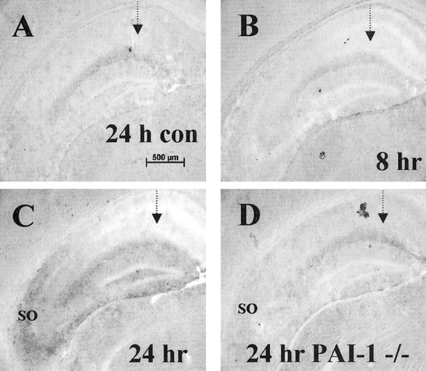
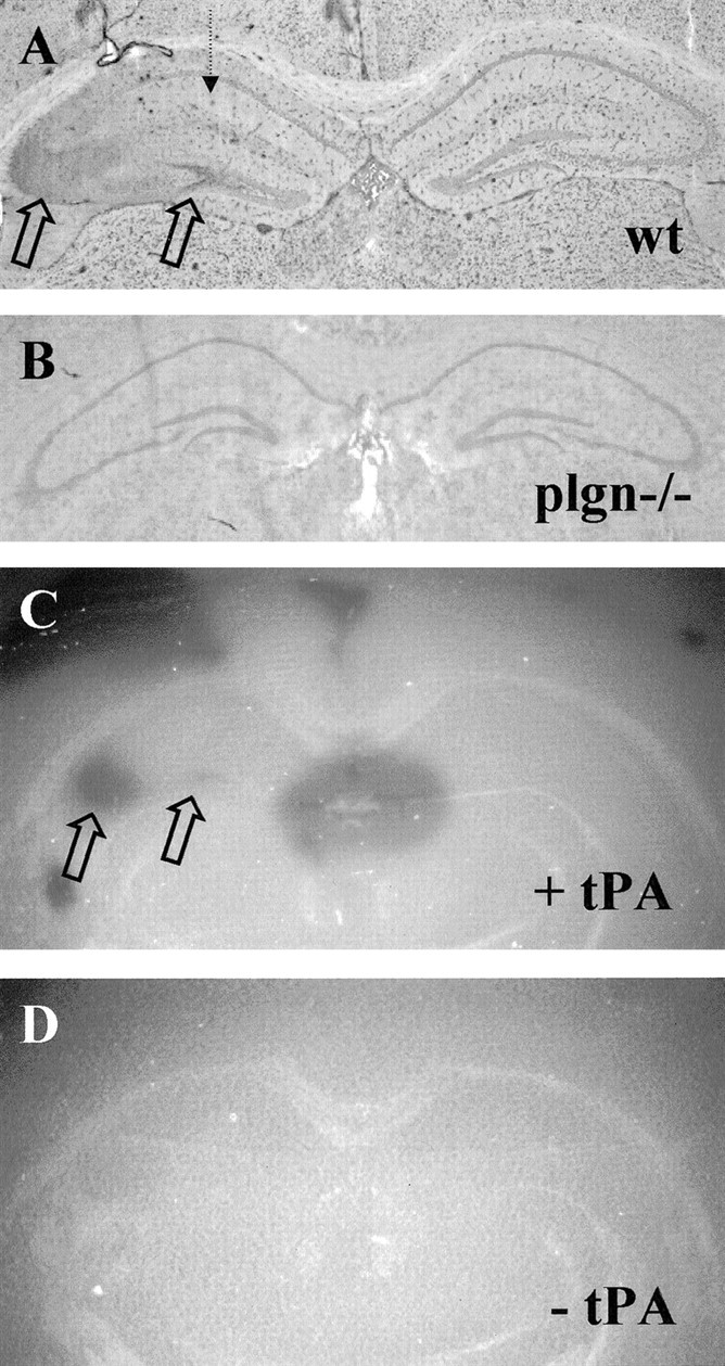
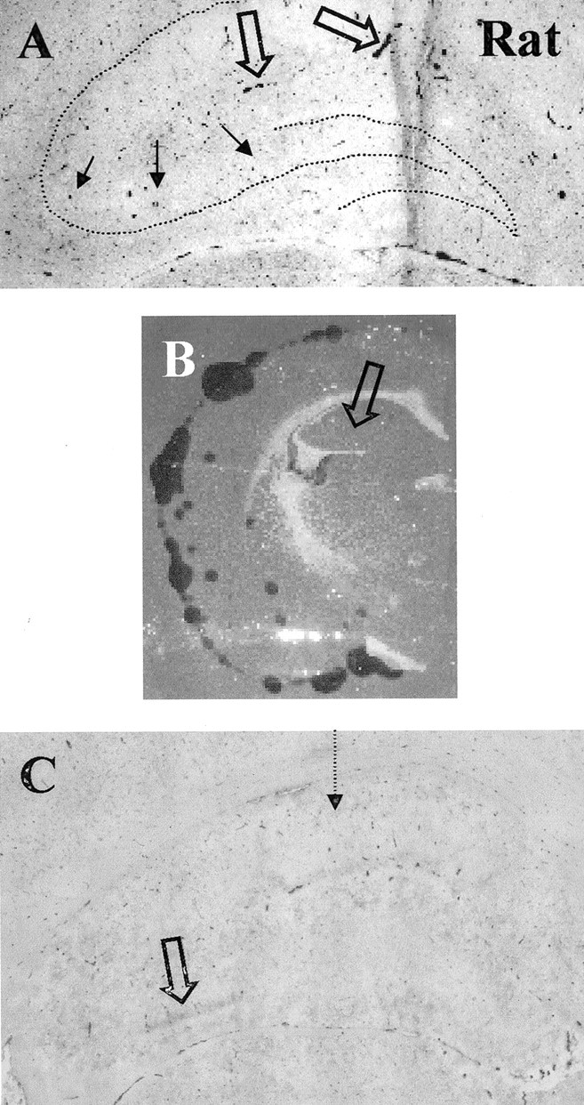
The extracellular protease cascade of tissue plasminogen activator (tPA) and plasminogen has been implicated in neuronal plasticity and degeneration. We show here that unstimulated expression of tPA in the mouse hippocampus is concentrated in the mossy fiber pathway, with little or no expression within the perforant path, the Schaffer collaterals, or neuronal cell bodies. tPA protein is also expressed in vascular endothelial cells throughout the brain parenchyma. Four hours after excitotoxic injury, tPA protein is transiently induced within CA1 pyramidal neurons. The induced CA1 tPA is localized to neurons that survive the injury and is enzymatically active. Within the mossy fiber pathway, injury resulted in decreased tPA protein. In contrast, mossy fiber tPA activity displayed a biphasic character: transient increase at 8 hr, then a decrease by 24 hr after injury. Analysis of plasminogen activator inhibitor-1 (PAI-1) expression showed that PAI-1 antigen is upregulated by 24 hr and could account for the tPA activity downregulation seen at this time point. Plasminogen immunohistochemistry suggested an increase within the mossy fiber pathway after injury. Finally, hippocampal tPA expression among various mammalian species was strikingly different. These results indicate a complex control of tPA protein and enzymatic activity in the hippocampus that may help regulate neuronal plasticity.


Figures

Fig. 1.
Fig. 1.
tPA is abundantly expressed in the mossy fiber pathway. A, B, tPA immunohistochemistry on wild-type (wt) sections shows robust tPA staining (red) in the hippocampal mossy fiber pathway. Sections are counter-stained with hematoxylin to visualize the neuronal cell body layers of the hippocampus. DG, Dentate gyrus;MF, mossy fiber pathway; n = 20 animals. C, D, tPA immunostain without the hematoxylin counterstain showing lack of significant staining within the neuronal cell bodies (compare high-magnification B withD). Arrows in D highlight vascular endothelial cell staining that is seen throughout the brain.E, F, tPA immunohistochemistry on wild-type sections after preabsorbing the antibody (Figure legend continues.) (Figure legend continued.) with tPA as described in Materials and Methods. Notice the loss of mossy fiber and vascular staining but residual background parenchymal stain;n = 7 animals. G, tPA immunohistochemistry on tPA-deficient (tPA−/−) brain sections. Notice a similar level of background parenchymal staining compared with E; n = 10 animals.H, Zymographic analysis of tPA activity (dark zone of lysis against a milky background) closely parallels staining seen by immunohistochemistry;n = 20 animals. Activity is plasminogen-dependent (data not shown). Boxes in A,C, and E show the region magnified inB, D, and F.Arrowheads in E–G highlight background CA2 cell body staining.
Fig. 2.
Fig. 2.
tPA is upregulated in CA1 neurons after excitotoxic injury. A, After a weak excitotoxic injury (between 0.3 and 0.6 nmol of KA in a 300 nl volume, depending on lot potency), at 8 hr after injury tPA protein is upregulated in neuronal cell bodies within the CA1 layer (arrow);n = 18 animals, with 14 showing some level of staining. These neurons occupy the border of surviving and degenerating neurons as seen in the adjacent section (B) stained with Nissl (arrow). Degenerating neurons are visualized as having pyknotic nuclei versus the surviving neurons that appear normal. C, After a robust injury (between 0.9 and 1.2 nmol of KA in a 300 nl volume, depending on lot potency), at 8 hr after injury tPA protein is strongly upregulated within some neuronal cell bodies of the contralateral uninjected hippocampus (arrows); n = 16 animals, with 8 showing significant staining. Inset, Magnification of several pyramidal neurons within the CA1 showing cytoplasmic tPA accumulation. Note that not all CA1 neurons upregulate tPA.D, This upregulated protein is active, as measured by the zymographic analysis. Contralateral CA1 tPA expression was seen when treated with sufficient KA to destroy ∼80% of ipsilateral hippocampus. In two animals, we could detect both ipsilateral and contralateral CA1 staining.
Fig. 3.
Fig. 3.
Time course of tPA induction in CA1 neurons after injury. Results show transiently upregulated neuronal cell body tPA in CA1 pyramidal neurons after injury in the contralateral uninjected hippocampus after a robust excitotoxic lesion. Sections were counterstained with hematoxylin to visualize the neuronal cell layer. Notice the lack of pyknotic nuclei and neuronal degeneration.A, No significant tPA staining is seen in the CA1 layer of control animals; n = 20 animals.B, CA1 neuronal cell layer 2.5 hr after lesion;n = 4 animals. C, tPA staining is induced 5 hr after injury (red staining within neuronal cell layer); n = 4 animals. D, tPA staining seen at 8 hr after injury; n = 8 animals.E, Forty-eight hours after injury, tPA is no longer detectable within the cell layer, yet the neurons still appear healthy;n = 4 animals. Additional time points assayed were 1 and 24 hr (data not shown).
Fig. 4.
Fig. 4.
tPA mRNA is strongly upregulated after excitotoxic injury in the contralateral uninjected CA1 pyramidal neurons byin situ hybridization. tPA mRNA was not detectable in control animals (n = 10; A) or tPA−/− animals (n = 10; C). tPA mRNA was only detectable after injury in wild-type animals (n = 5; B).
Fig. 5.
Fig. 5.
Modulation of protease inhibitor activity in the hippocampus after excitotoxic injury. The dashed arrowrepresents the site of excitotoxin injection. A, C, E, G, tPA protein staining; B, D, F, H, tPA activity zymographies performed on sections adjacent to those displayed for protein stains. A, B, Control PBS-injected wild type brain harvested 24 hr after injection; n = 6 animals. C, D, KA-injected brain harvested 8 hr after injection; n = 6 animals. E, F, KA-injected brain harvested 24 hr after injection;n = 6 animals. High-level upregulation of CA1 tPA protein was not seen in this experiment (see Fig. 2 legend). G, H, KA-injected brain from a PAI-1−/− animal harvested 24 hr after injection; n = 4 animals. All zymographies for each replicate shown were performed at the same time and incubated identically for the same time. The zymographies shown include the meningeal layer, which typically displays significant amounts of tPA activity, as an internal control to allow comparison of assay development time. The PAI-1 tissue showed slightly higher levels of tPA activity through out the tissue than wild type (H). Activities were all plasminogen-dependent. Open arrows indicate injected or ipsilateral hippocampus; closed arrows indicate uninjected or contralateral hippocampus.
Fig. 6.
Fig. 6.
PAI-1 upregulation in the hippocampus after excitotoxic injury. Ipsilateral injected hippocampus was immunostained for PAI-1. The dashed arrow represents the site of excitotoxin injection. A, Control PBS-injected wild-type brain harvested 24 hr after injection; n = 6 animals. B, KA-injected brain harvested 8 hr after injection; n = 6 animals. C, KA-injected brain harvested 24 hr after injection (SO, stratum oriens); n = 6 animals. D, KA-injected brain from a PAI-1−/− animal harvested 24 hr after injection; n = 4 animals.
Fig. 7.
Fig. 7.
Expression of plasminogen in the hippocampus after excitotoxic injury. The dashed arrow represents the site of excitotoxin injection. A, Plasminogen immunostain on KA-injected wild-type brain section 8 hr after injection. Open arrows highlight areas of intense staining;n = 6 animals. B, Plasminogen immunostain on plg−/− brain section; n = 3 animals. C, Plasminogen zymography of wild-type brain section adjacent to section shown in A indicating dark lytic zones of plasminogen activity (open arrows).D, Control zymography with tPA omitted from the mixture.
Fig. 8.
Fig. 8.
Rat mossy fiber pathway is devoid of significant tPA staining but is upregulated after excitotoxic injury.A, tPA protein is seen primarily in vascular endothelial cells. The dotted line shows the position of hippocampal neuronal cell layers. Filled arrows point to the mossy fiber pathway, where no significant tPA staining is detected.Open arrows point to tPA-stained vascular endothelium;n = 6 animals. B, In situ zymography assay showing tPA activity and lack of any detectable activity in the mossy fiber pathway. C, tPA protein staining is upregulated (open arrow) in a portion of the mossy fiber pathway 8 hr after stereotaxic KA injection into the CA1 region (dotted arrow);n = 8 animals, with 5 showing some level of tPA upregulation.

Similar articles

Cited by

References

    1. Akassoglou K, Kombrinck KW, Degen JL, Strickland S. Tissue plasminogen activator-mediated fibrinolysis protects against axonal degeneration and demyelination after sciatic nerve injury. J Cell Biol. 2000;149:1157–1166. - PMC - PubMed
    1. Andrade-Gordon P, Strickland S. Fractionation of heparin by chromatography on a tissue plasminogen activator-Sepharose column. Proc Natl Acad Sci USA. 1990;87:1865–1869. - PMC - PubMed
    1. Backstrom JR, Lim GP, Cullen MJ, Tökés ZA. Matrix metalloproteinase-9 (MMP-9) is synthesized in neurons of the human hippocampus and is capable of degrading the amyloid-β peptide (1–40). J Neurosci. 1996;16:7910–7919. - PMC - PubMed
    1. Baranes D, López-García JC, Chen M, Bailey CH, Kandel ER. Reconstitution of the hippocampal mossy fiber and associational-commissural pathways in a novel dissociated cell culture system. Proc Natl Acad Sci USA. 1996;93:4706–4711. - PMC - PubMed
    1. Baranes D, Lederfein D, Huang YY, Chen M, Bailey CH, Kandel ER. Tissue plasminogen activator contributes to the late phase of LTP and to synaptic growth in the hippocampal mossy fiber pathway. Neuron. 1998;21:813–825. - PubMed

Publication types

MeSH terms

LinkOut - more resources