. 2002 Mar 15;22(6):2142-52.
doi: 10.1523/JNEUROSCI.22-06-02142.2002.

Expression of a variant form of the glutamate transporter GLT1 in neuronal cultures and in neurons and astrocytes in the rat brain

Affiliations

Expression of a variant form of the glutamate transporter GLT1 in neuronal cultures and in neurons and astrocytes in the rat brain

Weizhi Chen et al. J Neurosci. .

Abstract

To identify glutamate transporters expressed in forebrain neurons, we prepared a cDNA library from rat forebrain neuronal cultures, previously shown to transport glutamate with high affinity and capacity. Using this library, we cloned two forms, varying in the C terminus, of the glutamate transporter GLT1. This transporter was previously found to be localized exclusively in astrocytes in the normal mature brain. Specific antibodies against the C-terminal peptides were used to show that forebrain neurons in culture express both GLT1a and GLT1b proteins. The pharmacological properties of glutamate transport mediated by GLT1a and GLT1b expressed in COS-7 cells and in neuronal cultures were indistinguishable. Both GLT1a and GLT1b were upregulated in astrocyte cultures by exposure to dibutyryl cAMP. We next investigated the expression of GLT1b in vivo. Northern blot analysis of forebrain RNA revealed two transcripts of approximately 3 and 11 kb that became more plentiful with developmental age. Immunoblot analysis showed high levels of expression in the cortex, hippocampus, striatum, thalamus, and midbrain. Pre-embedding electron microscopic immunocytochemistry with silver-enhanced immunogold detection was used to localize GLT1b in vivo. In the rat somatosensory cortex, GLT1b was clearly expressed in neurons in presynaptic terminals and dendritic shafts, as well as in astrocytes. The presence of GLT1b in neurons may offer a partial explanation for the observed uptake of glutamate by presynaptic terminals, for the preservation of input specificity at excitatory synapses, and may play a role in the pathophysiology of excitotoxicity.


Figures

Fig. 1.
Fig. 1.
Sequence comparison of variant forms of GLT1.A, The C-terminal sequence of the GLT1b clones obtained from a neuronal cDNA library is different from the published rat GLT1 (GLT1a) sequence U15098 (Roginski, 1996) in that the last 22 amino acids of GLT1a are replaced by a different stretch of 11 amino acids in GLT1b. B shows clones obtained by library screening and by PCR. RT-PCR was performed using the 5′ primer based on the known 5′-UTR GLT1 sequence and 3′ primers based on the 3′-UTR sequences of GLT1a and GLT1b cloned in this study. A 1.8 kb band was obtained with both pairs of primers and cloned into TOPO-TA vector (Invitrogen) and sequenced. Full-length clones were obtained with the structure shown that were subsequently used in expression studies. The base numbers used in this figure are according to the coding sequence.C shows the variant form GLT1b is formed by termination of the coding sequence after exon 9.
Fig. 2.
Fig. 2.
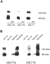
Immunoblot characterization of GLT1 antibodies.A, Membrane proteins from adult rat brain were analyzed by immunoblot as described (Materials and Methods) using anti-cGLT1a, anti-cGLT1b, and anti-nGLT1 antibodies (cGLT1a, cGLT1b, and nGLT1, respectively). All the anti-GLT1 antibodies recognized bands at ∼66 kDa and also at ∼130 kDa. Immunoreactivity was completely abolished when each antibody was preabsorbed with 50 μm of the peptide antigen (pep). Control experiments were done with heterologous peptides (anti-nGLT1 antibody preabsorbed with cGLT1a peptide, anti-cGLT1a antibody preabsorbed with cGLT1b peptide, and anti-cGLT1b antibody preabsorbed with cGLT1a), which had no effect on the immunoreactivity in brain membrane immunoblots. B, Immunoblot analysis showed that anti-cGLT1a and anti-cGLT1b antibodies have no cross-reactivity. Membrane proteins from cultured neurons, rat brain tissue, and COS-7 cells stably expressing GLT1a and GLT1b proteins were subjected to SDS-PAGE and immunoblotted with anti-cGLT1a and anti-cGLT1b antibodies. Anti-cGLT1a antibody (cGLT1a) recognized bands in GLT1a transfected COS-7 cells as well as in neuronal cultures and rat brain tissue, but not in GLT1b transfected COS-7 cells. Anti-cGLT1b antibody (cGLT1b) recognized bands in a GLT1b stable transfectant COS-7 cell line, in neuronal cultures and in rat brain membranes, but not in GLT1a-transfected cells.
Fig. 3.
Fig. 3.
Immunocytochemistry of GLT1a and GLT1b expression in cultured forebrain neurons. A, C,E, Cultured neurons at 18 d in vitroimmunostained with anti-cGLT1a (140 ng/ml) (A), anti-cGLT1b (3.2 μg/ml) (B), and anti-nGLT1 (1.5 μg/ml) (C) antibodies. All GLT1 antibodies detected immunoreactivity on neuronal processes and plasma membranes in a similar punctate pattern, suggesting synaptic localization of GLT1 proteins. Note that immunostaining by anti-cGLT1a and anti-cGLT1b antibodies was not distinguishable. B, D,F, Immunoreactivity was completely blocked using an excess of peptide against which each GLT1 antibody was directed.
Fig. 4.
Fig. 4.
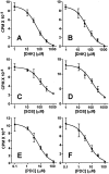
Effect of transport inhibitors on [3H]l-glutamate uptake in GLT1a- and GLT1b-transfected COS-7 cells. A,B, The effect of DHK on [3H]l-glutamate uptake by GLT1a (A) and GLT1b (B). In this particular experiment the IC50 values for GLT1a and GLT1b were 52 and 45 μm, respectively. C,D, The effect of SOS on [3H]l-glutamate uptake by GLT1a (C) and GLT1b (D). The IC50 values for GLT1a and GLT1b were 100 and 102 μm, respectively.E,F, The effect of PDC on [3H]l-glutamate uptake by GLT1a (E) and GLT1b (F). The IC50 values for GLT1a and GLT1b were 6.3 and 5.3 μm, respectively. In each case, results are single experiments representative of at least four experiments that were performed (Table 1).
Fig. 5.
Fig. 5.
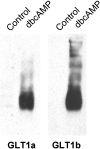
Similar upregulation of GLT1a and GLT1b in astrocytes in response to dibutyryl cAMP. Astrocyte cultures contained undetectable levels of GLT1a or GLT1b (Control). After treatment with 250 μm dibutyryl cAMP (dbcAMP) for 14 d, both GLT1a and GLT1b were significantly upregulated. Membrane protein (5 μg) solubilized in 1% SDS was loaded in each lane.
Fig. 6.
Fig. 6.
Expression of GLT1a and GLT1b mRNA during development. Total RNA was extracted from the forebrain of postnatal day 1 (P1), day 10 (P10), day 27 (P27), or adult (ad; 200 gm) Sprague Dawley rats. Ten micrograms of total RNA was loaded per lane. DNA probes representing 3′-UTR regions of GLT1a or GLT1b hybridized to RNA at ∼11 kb. The GLT1b probe clearly recognized another RNA ∼3 kb. At longer exposure times, a 3.5 kb band was seen with the GLT1a probe (data not shown). Expression of both GLT1a and GLT1b progressively increased through development.
Fig. 7.
Fig. 7.
Regional distribution of GLT1a and GLT1b proteins in rat brain. Immunoblots representing different rat brain regions were probed with anti-cGLT1a or anti-cGLT1b antibodies. Both proteins were detected in all regions. Strongest expression was observed in the cerebral cortex.
Fig. 8.
Fig. 8.
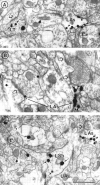
Anti-cGLT1b immunoreactivity in neurons and astrocytes of layer 1 of somatosensory cortex, as revealed by electron microscopic immunocytochemistry using SIG label. A, SIG label is present in an axon terminal (LT) and in an astrocyte (LAs). Here and in all other panels, SIG particles are indicated by arrows in neuronal elements and by arrowheads in astrocytes, whereas open arrowheads point to postsynaptic densities. SIG labeling is present along the plasma membrane of an astrocyte, including portions juxtaposed to asymmetric junctions (asterisk).B, An astrocyte is labeled LAs at two sites adjacent to two different asymmetric synapses (asterisks). C, A shaft, possessing a contiguous spine in the same section, is labeled LSh. An astrocyte is labeled LAs, and SIG particles are located adjacent to the plasma membrane. Note that not all astrocytes are labeled (e.g., C, UAs). Scale bar, 500 nm.

Similar articles

Cited by

References

    1. Aoki C, Venkatesan C, Go C-G, Mong JA, Dawson TM. Cellular and subcellular localization of NMDA-R1 subunit immunoreactivity in the visual cortex of adult and neonatal rats. J Neurosci. 1994;14:5202–5222. - PMC - PubMed
    1. Aoki C, Rodrigues S, Kurose H. Use of electron microscopy in the detection of adrenergic receptors. In: Machida CA, editor. Methods in molecular biology: adrenergic receptor protocols. Humana; Totowa, NJ: 1999. pp. 535–563. - PMC - PubMed
    1. Arriza JL, Fairman WA, Wadiche JI, Murdoch GH, Kavanaugh MP, Amara SG. Functional comparisons of three glutamate transporter subtypes cloned from human motor cortex. J Neurosci. 1994;14:5559–5569. - PMC - PubMed
    1. Arriza JL, Eliasof S, Kavanaugh MP, Amara SG. Excitatory amino acid transporter 5, a retinal glutamate transporter coupled to a chloride conductance. Proc Natl Acad Sci USA. 1997;94:4155–4160. - PMC - PubMed
    1. Asztely F, Erdemli G, Kullmann DM. Extrasynaptic glutamate spillover in the hippocampus: dependence on temperature and the role of active glutamate uptake. Neuron. 1997;18:281–293. - PubMed

Publication types

MeSH terms

Associated data

LinkOut - more resources