. 2002 Mar 15;22(6):2174-84.
doi: 10.1523/JNEUROSCI.22-06-02174.2002.

The mitochondrial toxin 3-nitropropionic acid induces striatal neurodegeneration via a c-Jun N-terminal kinase/c-Jun module

Affiliations

The mitochondrial toxin 3-nitropropionic acid induces striatal neurodegeneration via a c-Jun N-terminal kinase/c-Jun module

Marta Garcia et al. J Neurosci. .

Abstract

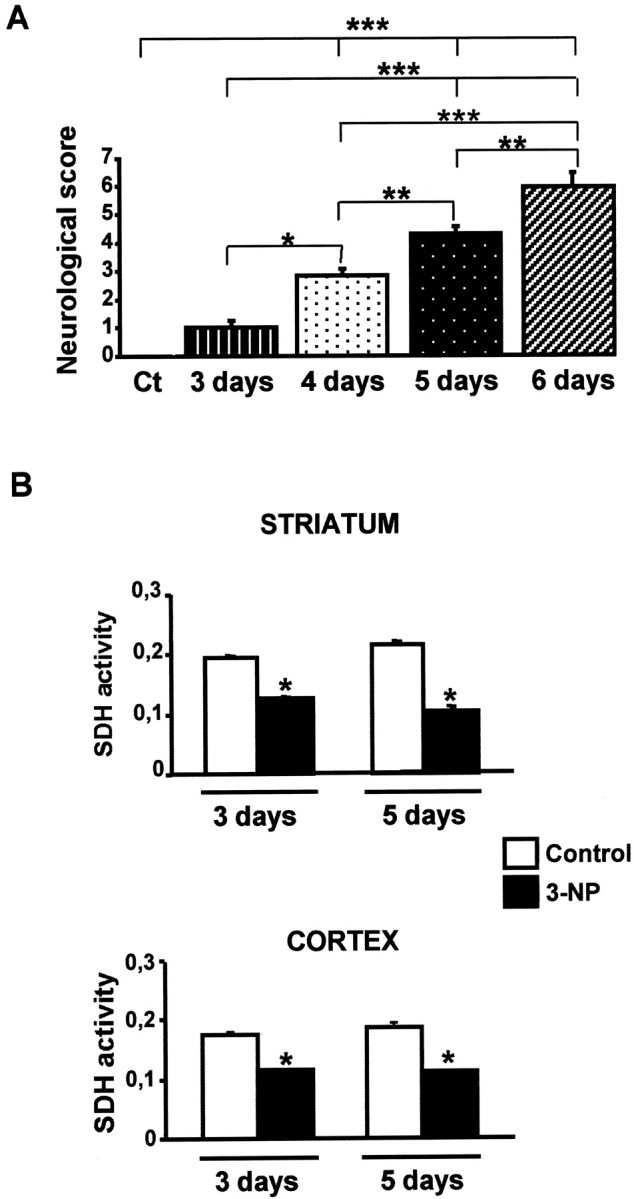
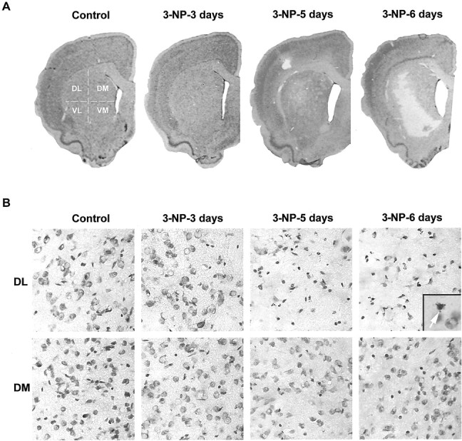
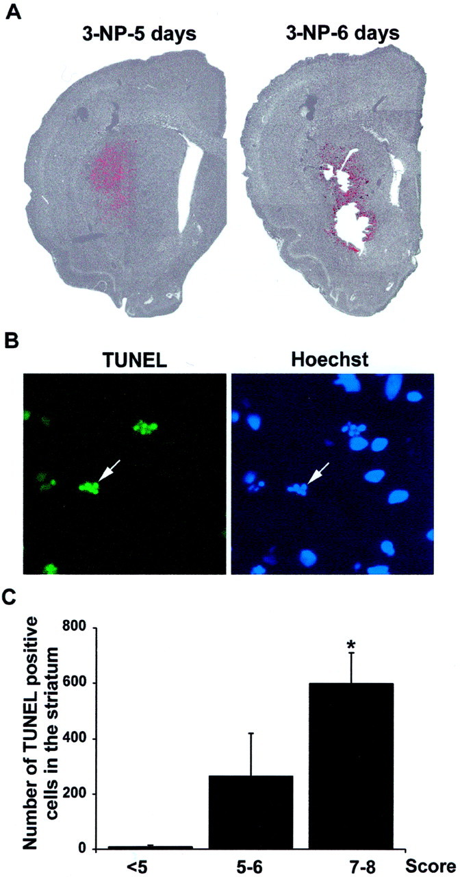
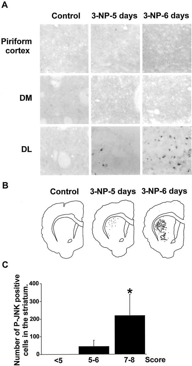
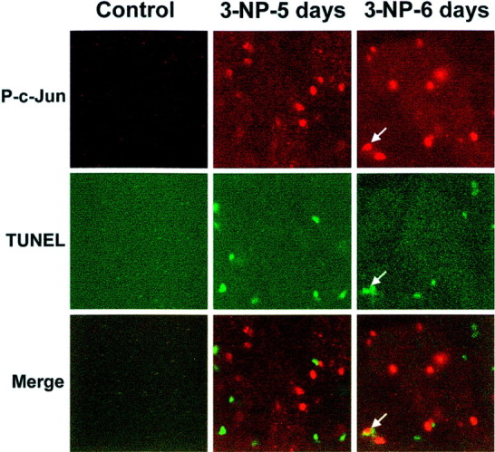
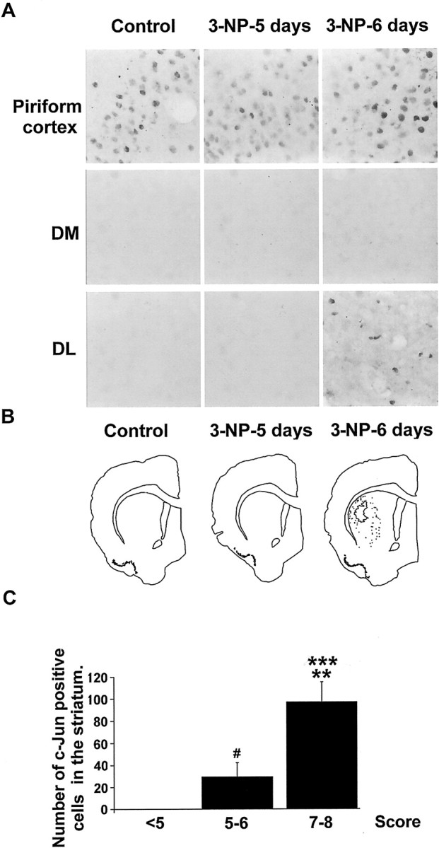
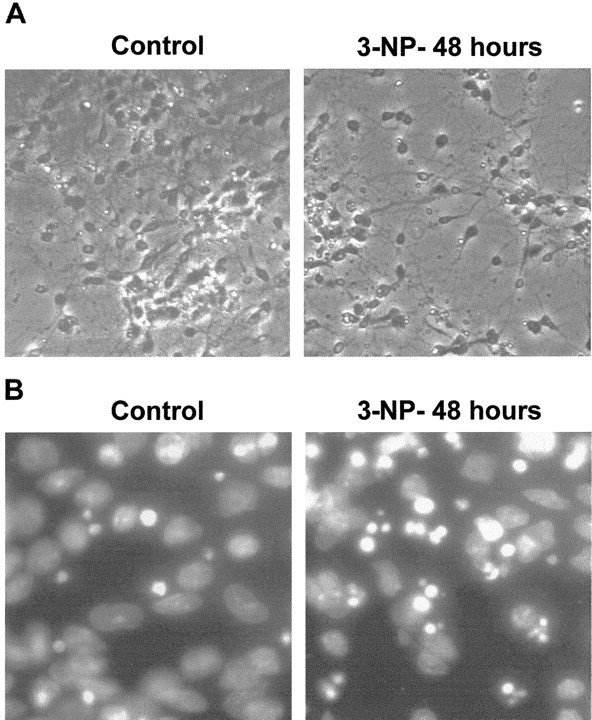
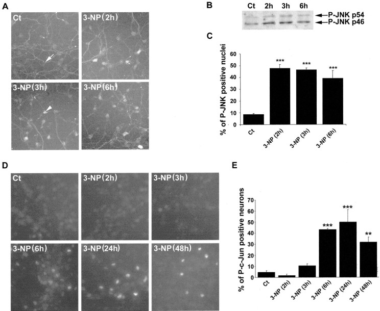
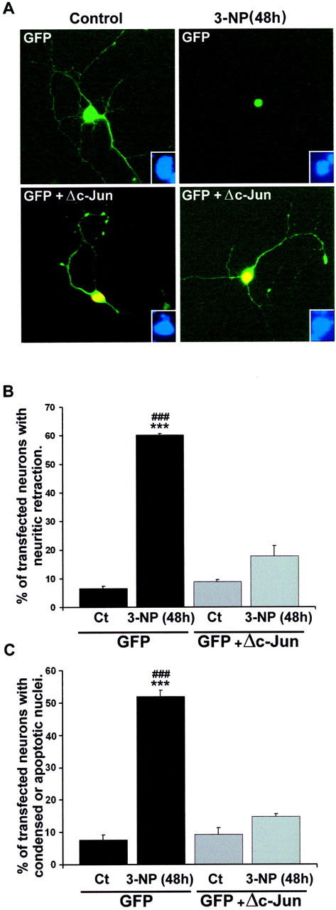
Impairments in mitochondrial energy metabolism are thought to be involved in most neurodegenerative diseases, including Huntington's disease (HD). Chronic administration of 3-nitropropionic acid (3-NP), a suicide inhibitor of succinate dehydrogenase, causes prolonged energy impairments and replicates most of the pathophysiological features of HD, including preferential striatal degeneration. In this study, we analyzed one of the mechanisms that could account for this selective 3-NP-induced striatal degeneration. In chronically 3-NP-infused rats, the time course of motor behavioral impairments and histological abnormalities was determined. Progressive alterations of motor performance occurred after 3 d. By histological analysis and terminal deoxynucleotidyl transferase-mediated biotinylated UTP nick end-labeling staining, we found a selective neurodegenerescence in the striatum, occurring first in its dorsolateral (DL) part. Activation of c-Jun N-terminal kinase (JNK) was analyzed from brain sections of these rats, using immunocytochemical detection of its phosphorylated form. Activation of JNK occurred progressively and selectively in the DL of the striatum and was followed by c-Jun activation and expression in the same striatal region. To elucidate the role of the JNK/c-Jun module in 3-NP-induced striatal degeneration, we then used primary striatal neurons in culture, in which we replicated neuronal death by application of 3-NP. We found strong nuclear translocation of activated JNK that was rapidly followed by phosphorylation of the transcription factor c-Jun. Overexpression of a dominant negative version of c-Jun, lacking its transactivation domain and phosphorylation sites for activated JNK, completely abolished 3-NP-induced striatal neurodegeneration. We thus conclude that a genetic program controlled by the JNK/c-Jun module is an important molecular event in 3-NP-induced striatal degeneration.


Figures

Fig. 1.
Fig. 1.
Systemic administration of 3-NP leads to neurological impairment and inhibition of SDH. A, Control (Ct) and 3-NP-treated rats were tested every day for behavioral performance (from day 3). Score 1 corresponded to mild and intermittent dystonia of one hindlimb, scores 2–3, intermittent or permanent appearance of dystonia for two hindlimbs; scores 4–5, wobbling gait and lack of grasping; and scores 6–8, severe dystonic posturing to recumbency. Data are representative of at least six rats per group. Statistical analysis: *p < 0.05; **p < 0.005; ***p < 0.001 when comparing control and 3-NP-treated rats (Scheffé test).B, Striatal and cortical SDH activity was measured by semiquantitative immunohistochemistry. Note the strong decrease in both cortical and striatal slices. *p < 0.001 when comparing 3-NP-treated rats with their corresponding control (Scheffé test).
Fig. 2.
Fig. 2.
Systemic administration of 3-NP leads to a progressive and selective degeneration in the striatum. Cresyl violet staining was performed on brain sections from control and 3-NP-treated rats. A, Low magnification showing the selective loss of cells in the striatum at day 5. B, High magnification of cresyl violet staining in the DL and DM parts of the striatum. Note the progressive cell loss in the DL striatum on 3-NP treatment when compared with the DM striatum.Inset, Fragmented nucleus displaying apoptotic bodies.VL, Ventrolateral; VM, ventromedial.
Fig. 3.
Fig. 3.
TUNEL immunoreactivity correlates with neurological scores. A, Low magnification showing TUNEL-reactive cells (revealed by FITC and depicted here inred) superimposed with Hoechst staining (showed here ingray) in the striatum. Note the strong TUNEL labeling that appears at 3-NP-5 days in the DL striatum.B, Higher magnification of TUNEL-positive cells showing apoptotic bodies (arrow) at day 5 of the 3-NP treatment showing fragmented nuclei (arrow). Right panel, Corresponding Hoechst staining. C, TUNEL-positive cells were counted in the striatum. The mean value was determined in group of 3-NP-treated rats presenting the same neurological score. <5, Three to 4 d of 3-NP treatment; 5–6, 5 d of treatment;7–8, 6 d of treatment. Statistical analysis: *p < 0.05 when comparing scores7–8 and <5 (Scheffé test).
Fig. 4.
Fig. 4.
Systemic administration of 3-NP leads to a progressive activation of JNK in the striatum selectively. JNK activation was analyzed by immunocytochemical detection of its active, phosphorylated form (P-JNK) from sections corresponding to the rats used for behavioral, histological, and TUNEL analysis (see previous figures). A, High magnification of P-JNK immunoreactivity in the piriform cortex and DM and DL regions of the striatum. Note the lack of labeling in the Piriform cortex and the DM striatum inControl and 3-NP-treated rats. P-JNK immunoreactivity appears at 3-NP-5 days in theDL striatum specifically. B, P-JNK-immunoreactive cells were depicted in the striatum using an image analyzer. Note their appearance in the DL striatum specifically at 3-NP-5 days treatment. C, Quantification of P-JNK-immunoreactive cells was performed from at least five different rats per neurological score. For each rat, one equivalent striatal section (30 μm) was counted. Shown is the mean value determined in 3-NP-treated rats presenting the same neurological score. <5, Three to 4 d of 3-NP treatment;5–6, 5 d of treatment; 7–8, 6 d of treatment. Statistical analysis: *p < 0.005 when comparing scores 7–8 and <5(Scheffé test).
Fig. 5.
Fig. 5.
Activation of c-Jun phosphorylation occurs in the degenerative striatal region. P-c-Jun immunoreactivity was performed using an anti-P-c-Jun antibody. Note a strong induction at3-NP-5 days and 3-NP-6 days treatment. Coimmunostaining with TUNEL indicates that P-c-Jun immunoreactivity occurs in the degenerative region. Note the double labeling between P-c-Jun and TUNEL staining in one striatal neuron (bottom right panel, arrow).
Fig. 6.
Fig. 6.
Systemic administration of 3-NP leads to a progressive expression of c-Jun in the striatum, selectively. c-Jun expression was analyzed by immunocytochemistry using a selective antibody. A, High magnification of c-Jun immunoreactivity in the Piriform cortex shows basal expression in this region in both Control and3-NP-treated rats. Note that c-Jun expression occurs in the DL part of the striatum at 3-NP-6 days treatment. B, c-Jun-immunoreactive cells were depicted in the striatum using an image analyzer. Note their appearance in the DL striatum specifically at 3-NP-6 days treatment. C, Quantification of c-Jun-immunoreactive cells was performed as in Figure 4. Statistical analysis: **p < 0.01 when comparing scores5–6 and 7–8; ***p< 0.0001 when comparing scores 7–8 and <5; #p < 0.05 when comparing scores 5–6 and <5(Scheffé test).
Fig. 7.
Fig. 7.
3-NP leads to apoptosis in primary striatal cultures. High magnification is shown of phase contrast (A) and Hoechst staining (B) from primary striatal cultures treated with 3-NP (1 mm) for 48 hr (3-NP-48 hours).Control, Untreated striatal neurons.
Fig. 8.
Fig. 8.
3-NP leads to activation of the JNK/c-Jun module in cultures of primary striatal neurons. A, P-JNK immunoreactivity in control (Ct) and 3-NP-treated cells. Note the low P-JNK immunoreactivity in control cells restricted to the neuritic extension (white arrow). Note also the strong nuclear translocation of P-JNK immunoreactivity after3-NP(3h) treatment (arrowhead).B, Western blot analysis of P-JNK immunoreactivity.C, Quantification of P-JNK-immunoreactive nuclei was performed and compared with the total number of striatal neurons (analyzed by Hoechst staining). Data are representative of three independent experiments (for each experiment, the mean value was calculated from 5 randomly chosen fields, representing 50 neurons each). Statistical analysis: ***p < 0.001 when comparing P-JNK-immunoreactive nuclei between3-NP-treated and control (Ct) neurons (Scheffé test). D, Phosphorylation of c-Jun was determined using an antibody specific for its phosphorylated form (P-c-Jun). Note that P-c-Jun-immunoreactive neurons appear at3-NP(3h) treatment. E, Quantification of P-c-Jun-positive nuclei was performed as detailed for P-JNK. Statistical analysis: **p < 0.001 and ***p < 0.0001 when comparing control (Ct) and 3-NP-treated neurons (Scheffé test).
Fig. 9.
Fig. 9.
A dominant negative form of c-Jun protects neurons from apoptosis induced by 3-NP. Primary striatal neurons were transfected with GFP alone or in combination with a dominant negative form of c-Jun (Δc-Jun, lacking the first 169 amino acids). A, Transfected neurons were visualized by GFP (top panels, green). In cotransfected neurons (bottom panels), detection of Δc-Jun expression was performed using an antibody that recognizes the FLAG epitope. The coexpression of GFP and Δc-Jun is shown in yellow. Note the protection from 3-NP-induced neuritic retraction and nuclear condensation in cells cotransfected with GFP and Δc-Jun.Insets, Corresponding Hoechst staining in these neurons.B, Quantification of neurite retraction from transfected cells. C, Quantification of transfected neurons having condensed or fragmented nuclei. Data are representative of three independent experiments (for each experiment, ∼500 transfected neurons were analyzed). Statistical analysis ***p< 0.001 when comparing controls (Ct) with3-NP treatment; ###p < 0.001 when comparing 3-NP-treated neurons transfected with GFP alone or withGFPc-Jun (Scheffé test).

Similar articles

Cited by

References

    1. Angel P, Hattori K, Smeal T, Karin M. The jun proto-oncogene is positively autoregulated by its product, Jun/AP-1. Cell. 1988;55:875–885. - PubMed
    1. Beal MF. Mitochondria, free radicals, and neurodegeneration. Curr Opin Neurobiol. 1996;6:661–666. - PubMed
    1. Beal MF. Energetics in the pathogenesis of neurodegenerative diseases. Trends Neurosci. 2000;23:298–304. - PubMed
    1. Beal MF, Brouillet E, Jenkins BG, Ferrante RJ, Kowall NW, Miller JM, Storey E, Srivastava R, Rosen BR, Hyman BT. Neurochemical and histologic characterization of striatal excitotoxic lesions produced by the mitochondrial toxin 3-nitropropionic acid. J Neurosci. 1993;13:4181–4192. - PMC - PubMed
    1. Behrens A, Sibilia M, Wagner EF. Amino-terminal phosphorylation of c-Jun regulates stress-induced apoptosis and cellular proliferation. Nat Genet. 1999;21:326–329. - PubMed

Publication types

MeSH terms