

Endosomal compartments serve multiple hippocampal dendritic spines from a widespread rather than a local store of recycling membrane
- PMID: 11896161
- PMCID: PMC6758269
- DOI: 10.1523/JNEUROSCI.22-06-02215.2002
Endosomal compartments serve multiple hippocampal dendritic spines from a widespread rather than a local store of recycling membrane
Abstract
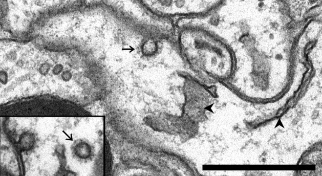
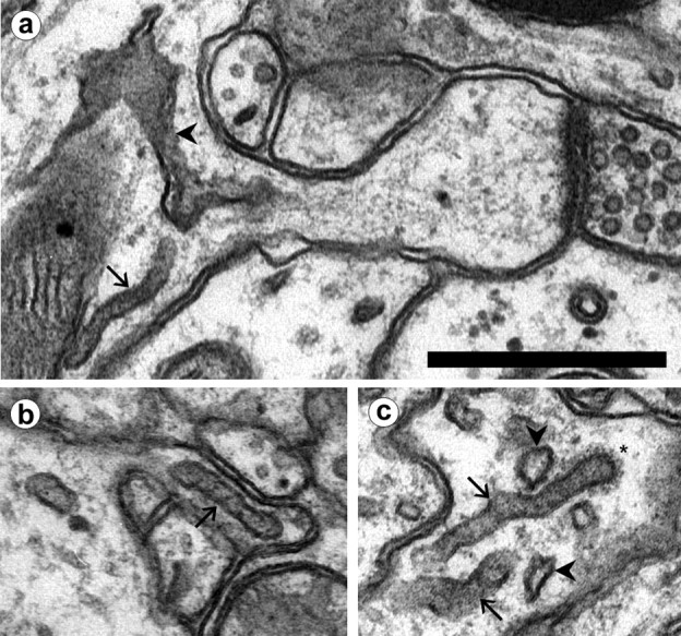
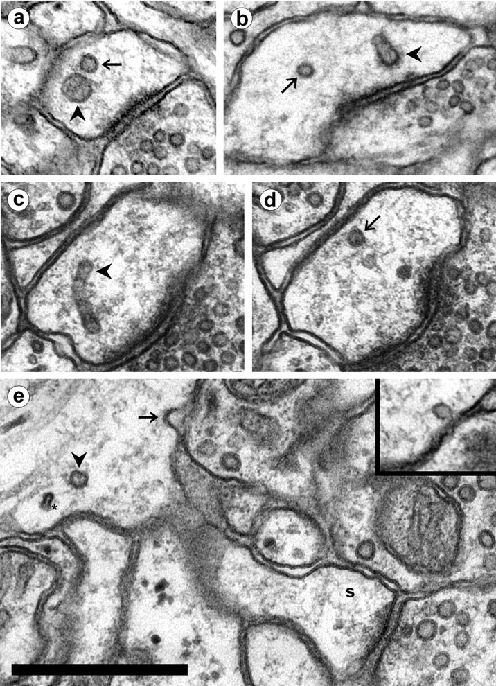
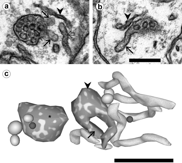
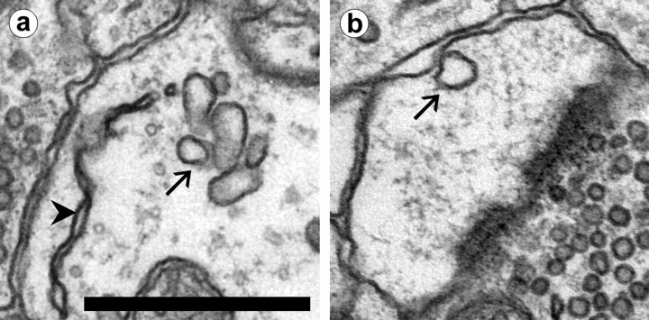
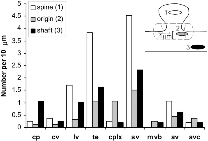
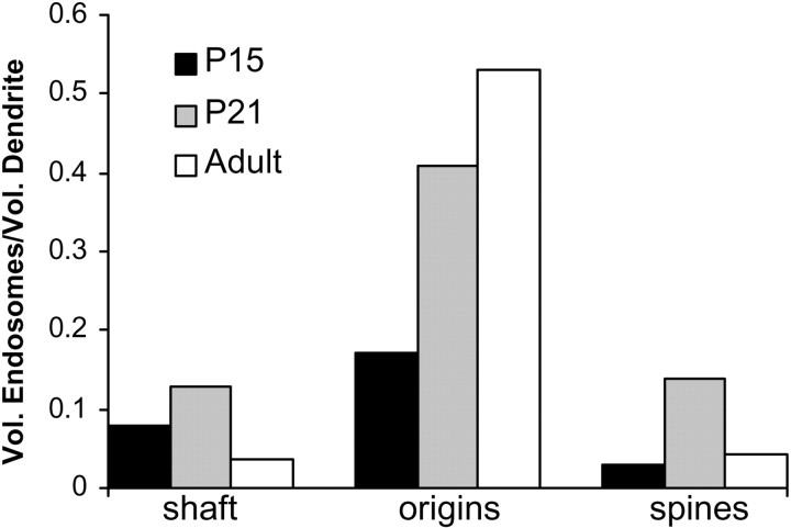
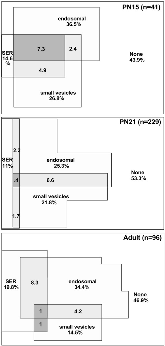
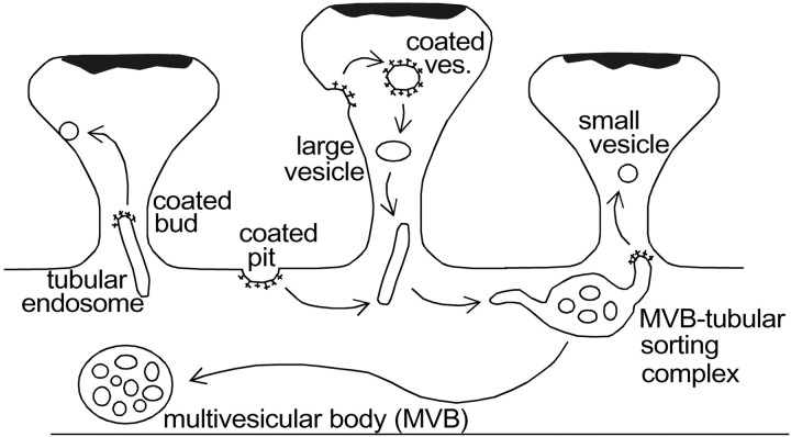
Endosomes are essential to dendritic and synaptic function in sorting membrane proteins for degradation or recycling, yet little is known about their locations near synapses. Here, serial electron microscopy was used to ascertain the morphology and distribution of all membranous intracellular compartments in distal dendrites of hippocampal CA1 pyramidal neurons in juvenile and adult rats. First, the continuous network of smooth endoplasmic reticulum (SER) was traced throughout dendritic segments and their spines. SER occupied the cortex of the dendritic shaft and extended into 14% of spines. Several types of non-SER compartments were then identified, including clathrin-coated vesicles and pits, large uncoated vesicles, tubular compartments, multivesicular bodies (MVBs), and MVB-tubule complexes. The uptake of extracellular gold particles indicated that these compartments were endosomal in origin. Small, round vesicles and pits that did not contain gold were also identified. The tubular compartments exhibited clathrin-coated tips consistent with the genesis of these small, presumably exosomal vesicles. Approximately 70% of the non-SER compartments were located within or at the base of dendritic spines. Overall, only 29% of dendritic spines had endosomal compartments, whereas 20% contained small vesicles. Small vesicles did not colocalize in spines with endosomes or SER. Three-dimensional reconstructions revealed that up to 20 spines shared a recycling pool of plasmalemmal proteins rather than maintaining independent stores at each spine.
Figures


Similar articles
-
Three-dimensional organization of smooth endoplasmic reticulum in hippocampal CA1 dendrites and dendritic spines of the immature and mature rat.J Neurosci. 1997 Jan 1;17(1):190-203. doi: 10.1523/JNEUROSCI.17-01-00190.1997. J Neurosci. 1997. PMID: 8987748 Free PMC article.
-
Trans-endocytosis via spinules in adult rat hippocampus.J Neurosci. 2004 Apr 28;24(17):4233-41. doi: 10.1523/JNEUROSCI.0287-04.2004. J Neurosci. 2004. PMID: 15115819 Free PMC article.
-
Three-dimensional analysis of the structure and composition of CA3 branched dendritic spines and their synaptic relationships with mossy fiber boutons in the rat hippocampus.J Comp Neurol. 1992 Nov 8;325(2):169-82. doi: 10.1002/cne.903250204. J Comp Neurol. 1992. PMID: 1460112
-
The role of endosomal-recycling in long-term potentiation.Cell Mol Life Sci. 2011 Jan;68(2):185-94. doi: 10.1007/s00018-010-0516-2. Epub 2010 Sep 6. Cell Mol Life Sci. 2011. PMID: 20820847 Free PMC article. Review.
-
[Three-dimentional organization of synapses and astroglia in the hippocampus of rats and ground squirrels: new structural and functional paradigms of the synapse function].Biofizika. 2003 Mar-Apr;48(2):289-308. Biofizika. 2003. PMID: 12723356 Review. Russian.
Cited by
-
Phosphatidylinositol-3-phosphate mediates Arc capsid secretion through the multivesicular body pathway.Proc Natl Acad Sci U S A. 2024 Aug 27;121(35):e2322422121. doi: 10.1073/pnas.2322422121. Epub 2024 Aug 23. Proc Natl Acad Sci U S A. 2024. PMID: 39178227 Free PMC article.
-
The ataxia (axJ) mutation causes abnormal GABAA receptor turnover in mice.PLoS Genet. 2009 Sep;5(9):e1000631. doi: 10.1371/journal.pgen.1000631. Epub 2009 Sep 4. PLoS Genet. 2009. PMID: 19759851 Free PMC article.
-
The F-BAR protein Rapostlin regulates dendritic spine formation in hippocampal neurons.J Biol Chem. 2011 Sep 16;286(37):32672-83. doi: 10.1074/jbc.M111.236265. Epub 2011 Jul 15. J Biol Chem. 2011. PMID: 21768103 Free PMC article.
-
Golgi-independent secretory trafficking through recycling endosomes in neuronal dendrites and spines.Elife. 2017 Sep 6;6:e27362. doi: 10.7554/eLife.27362. Elife. 2017. PMID: 28875935 Free PMC article.
-
Plasticity-induced growth of dendritic spines by exocytic trafficking from recycling endosomes.Neuron. 2006 Dec 7;52(5):817-30. doi: 10.1016/j.neuron.2006.09.040. Neuron. 2006. PMID: 17145503 Free PMC article.
References
-
- Bi G, Poo M. Synaptic modification by correlated activity: Hebb's postulate revisited. Annu Rev Neurosci. 2001;24:139–166. - PubMed
-
- Buchs PA, Stoppini L, Párducz A, Siklós L, Muller D. A new cytochemical method for the ultrastructural localization of calcium in the central nervous system. J Neurosci Methods. 1994;54:83–93. - PubMed
-
- Carroll RC, Beattie EC, von Zastrow M, Malenka RC. Role of AMPA receptor endocytosis in synaptic plasticity. Nat Rev Neurosci. 2001;2:315–324. - PubMed
Publication types
MeSH terms
Substances
Grants and funding
LinkOut - more resources
Full Text Sources
Miscellaneous