. 2002 Mar 15;22(6):2225-36.
doi: 10.1523/JNEUROSCI.22-06-02225.2002.

Versican is upregulated in CNS injury and is a product of oligodendrocyte lineage cells

Affiliations

Versican is upregulated in CNS injury and is a product of oligodendrocyte lineage cells

Richard A Asher et al. J Neurosci. .

Abstract

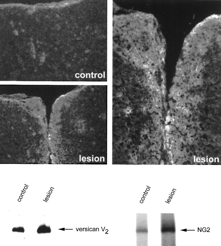
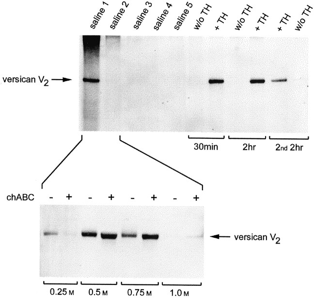
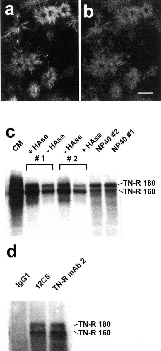
Chondroitin sulfate proteoglycan (CS-PG) expression is increased in response to CNS injury and limits the capacity for axonal regeneration. Previously we have shown that neurocan is one of the CS-PGs that is upregulated (Asher et al., 2000). Here we show that another member of the aggrecan family, versican, is also upregulated in response to CNS injury. Labeling of frozen sections 7 d after a unilateral knife lesion to the cerebral cortex revealed a clear increase in versican immunoreactivity around the lesion. Western blot analysis of extracts prepared from injured and uninjured tissue also revealed considerably more versican in the injured tissue extract. In vitro studies revealed versican to be a product of oligodendrocyte lineage cells (OLCs). Labeling was seen between the late A2B5-positive stage and the O1-positive pre-oligodendrocyte stage. Neither immature, bipolar A2B5-positive cells, nor differentiated, myelin-forming oligodendrocytes were labeled. The amount of versican in conditioned medium increased as these cells differentiated. Versican and tenascin-R colocalized in OLCs, and coimmunoprecipitation indicated that the two exist as a complex in oligodendrocyte-conditioned medium. Treatment of pre-oligodendrocytes with hyaluronidase led to the release of versican, indicating that its retention at the cell surface is dependent on hyaluronate (HA). In rat brain, approximately half of the versican is bound to hyaluronate. We also provide evidence of a role for CS-PGs in the axon growth-inhibitory properties of oligodendrocytes. Because large numbers of OLCs are recruited to CNS lesions, these results suggest that OLC-derived versican contributes to the inhospitable environment of the injured CNS.


Figures

Fig. 1.
Fig. 1.
Versican is upregulated in the injured CNS.Top, Immunolocalization of versican in the injured CNS. Coronal frozen sections were labeled with the anti-versican mAb 12C5 7 d after a knife cut lesion to the cerebral cortex. The dorsal surface of the brain is topmost. Labeling is apparent around the injury (lesion), which is clearly lacking on the uninjured side (control).Bottom, Western blot analysis of versican in the injured brain. SDS extracts were prepared from injured and uninjured cerebral cortex. The extracts were equalized for total protein (162 μg), run in a 4% gel under nonreducing conditions, and transferred to polyvinylidene difluoride. The blot was labeled with the anti-versican mAb 12C5 and then with rabbit antibodies against the NG2 proteoglycan. An upregulation of both CS-PGs was clearly evident in the injured brain extracts.
Fig. 2.
Fig. 2.
Pro-oligodendroblasts and pre-oligodendrocytes, but not bipolar oligodendrocyte precursor cells or myelin-forming oligodendrocytes, label for versican. OPCs were grown for 1 d (a–c), 2 d (d–f), or 5 d (j–l) in differentiation medium, or in division medium for 2 d and then in differentiation medium for 2 d (g–i). The cells were double labeled for A2B5 (a) or O1 (d, g, j) and versican (b, e, h, k). The two are shown together in theright-hand column (c, f, i, l). The labeling was performed on living cells, and the cells were post-fixed in cold methanol. In OPCs plated directly in differentiation medium, versican is first seen to be associated with weakly A2B5-positive, multipolar cells (ac,arrowheads) and subsequently with O1-positive cells (d–f). In OPCs grown initially in medium containing PDGF and FGF2 and then in differentiation medium, versican appears as a ring on the dorsal (top) cell surface (g–i). Neither bipolar, A2B5-positive OPCs (ac, arrows) nor myelin-forming oligodendrocytes (j–l) label for versican. Scale bars, 20 μm.
Fig. 3.
Fig. 3.
Oligodendrocytes produce the same versican isoform (V2) as that found in the rat CNS. The versican in adult rat brain (the 0.5 m NaCl fraction from DEAE-bound material; see Fig. 5) was compared with that in the conditioned medium of OLCs, astrocytes, meningeal cells, and the OPC-like cell line CG4. A testicular hyaluronidase (test. hyal.) extract of rat brain was overloaded to show that low levels of V0 and V1 are present in adult rat brain. OLCs make only the V2 isoform, which is unable to enter the gel without chondroitinase treatment. Astrocytes do not produce versican. Meningeal cells make only V0 and V1, both of which require chondroitinase digestion to enter the gel. As expected, CG4 cells make mostly V2, although small amounts of V0 and V1 were also detected.
Fig. 4.
Fig. 4.
Versican binding to pre-oligodendrocytes is hyaluronate-dependent. Pre-oligodendrocytes were double labeled for O1 and versican (a, b), hyaluronate and versican (c, d), and O1 and versican (e, f). The cells shown in e and f were digested withStreptomyces hyaluronidase before labeling. Versican labeling takes the form of a ring, wholly or partly encircling the cell body of O1-positive pre-oligodendrocytes (b, d). The distribution of hyaluronate is identical to that of versican (c, d). Strepto-myces hyaluronidase abolished versican labeling (f, shows Hoechst-labeled nuclei). Scale bar, 20 μm. g, Two 25 cm2 flasks of OPCs were grown for 2 d in medium containing PDGF and FGF2 (div). This medium was then changed to one supportive of oligodendrocyte differentiation (diff), and the cells were grown for a further 24 hr. This medium was then removed and replaced with the same medium, either with (#1) or without (#2) testicular hyaluronidase (50 μg/ml) for 1 hr. This medium was removed and replaced with the same medium, except that the enzyme was added to the flask not previously treated with hyaluronidase (#2), but not to flask #1. The conditioned media were concentrated and treated with chondroitinase ABC. An equal volume of each was run under nonreducing conditions in a 4% gel (forversican and neurocan) or under reducing conditions in a 7% gel (brevican) and transferred to nitrocellulose. The blots were labeled with the anti-versican mAb 12C5, the anti-neurocan mAb 1G2, or an anti-brevican mAb. Versican, but not neurocan or brevican, was released intact from pre-oligodendrocytes by hyaluronidase. The amount of versican detected in the conditioned medium of differentiating OLCs was greater than that in dividing cells. This was not the case for either neurocan or brevican.
Fig. 5.
Fig. 5.
Versican can be released from adult rat brain with hyaluronidase. Top, Serial extracts were prepared from adult rat brain with PBS, pH 5.3 (saline 1–5). After the fifth PBS extract, the homogenate was divided into two equal parts, to only one of which was added testicular hyaluronidase (TH). The homogenates were incubated at 37°C for 2 hr. An aliquot was removed from each after 30 min. A second 2 hr incubation was then set up, in which hyaluronidase was added to the homogenate not previously exposed to the enzyme. All extracts were treated with chondroitinase ABC. The volume of each extract was adjusted according to the amount of tissue from which it derived, run in a 4% gel under nonreducing conditions, and transferred to nitrocellulose. The blot was labeled with the anti-versican mAb 12C5. Versican was present in the first saline extract, but the release of additional amounts required hyaluronidase. Bottom, The first and second saline extracts were pooled and DEAE cellulose was added. The bound proteins were eluted with increasing concentrations of NaCl. Versican (V2) bound to the anion exchange resin and was eluted between 0.5 and 0.75 msalt. Chondroitinase brought about a small but discrete shift, indicating that versican V2 carries little chondroitin sulfate.
Fig. 6.
Fig. 6.
Versican colocalizes with tenascin-R, and the two exist as a complex in oligodendrocyte-conditioned medium. OPCs were grown for 1 d in differentiation medium and then double labeled with the 12C5 anti-versican mAb (a) and a polyclonal (rabbit) anti-tenascin-R (b). The distributions of the two appear identical. Scale bar, 25 μm.c, Two flasks of OPCs were grown for 2 d in differentiation medium. This was then removed and replaced with the same medium, either with (#1) or without (#2) testicular hyaluronidase (50 μg/ml) for 1 hr. This medium was removed and replaced with the same medium, except that the enzyme was added to the flask not previously treated with the enzyme (#2), but not to flask #1. The conditioned media were concentrated and equalized according to the protein content of the (NP-40) cell lysate. The samples were run under reducing conditions in a 7% gel, transferred to nitrocellulose, and labeled with an anti-tenascin-R mAb. Large amounts of tenascin-R were detected in the conditioned medium of oligodendrocytes (CM). Although tenascin-R was present in the hyaluronidase-treated cells (#1 +HAse), the amount was no greater than that present in the conditioned medium of the untreated cells (#2 −HAse). The failure of hyaluronidase to release tenascin-R is also evidenced by the presence of tenascin-R in the cell lysates (NP40). Note that the smaller tenascin-R 160 predominates in the conditioned media, whereas the 180 kDa form predominates in the cell lysates. d, Immunoprecipitation was performed on oligodendrocyte-conditioned medium with an isotype-matched control antibody (IgG1), the anti-versican mAb 12C5, or an anti-tenascin-R mAb. The products were run in a 7% gel under reducing conditions, transferred to nitrocellulose, and labeled with the same anti-tenascin-R mAb. The anti-versican mAb immunoprecipitated tenascin-R, indicating that the two are physically associated in oligodendrocyte-conditioned medium.
Fig. 7.
Fig. 7.
Versican is upregulated by TGFβ in dividing OPCs and by IL-1β and CNTF in differentiating oligodendrocytes. Dividing cells were maintained for 2 d in medium containing PDGF and FGF2 and the cytokine/growth factor (10 ng/ml) being tested (a, b). Differentiating cells were grown for 2 d in medium containing PDGF and FGF2, and then for a further 2 d in differentiation medium containing the cytokine/growth factor (10 ng/ml) under investigation (c). The conditioned media were collected, concentrated, and treated with chondroitinase ABC. The samples were equalized according to the protein content of the cell lysates, run under nonreducing conditions in a 4% gel, and transferred to nitrocellulose. The blots were labeled with the anti-versican mAb 12C5. TGFβ led to an increase in the amount of versican present in the conditioned media of dividing OPCs (a, b). In differentiating cells, IL-1β and CNTF, but not TGFβ, brought about an increase in the amount of versican in the conditioned medium (c).
Fig. 8.
Fig. 8.
Chondroitin sulfate proteoglycans contribute to the axon growth-inhibitory properties of oligodendrocytes. Dorsal root ganglion neurons growing on untreated (a, c) and chondroitinase ABC-treated (b, d) paraformaldehyde-fixed pro-oligodendroblasts were labeled with 3A10 (a, b) and Hoechst 33342 to visualize nuclei (c, d). Scale bar, 100 μm. e, Quantification of neurite outgrowth on pro-oligodendroblasts. Chondroitinase treatment led to a significant increase in DRG neurite outgrowth (**p < 0.001; Student's t test) without affecting neuronal adhesion to the substrate. These findings suggest that axon growth in an oligodendroglial environment is impaired by chondroitin sulfate.

Similar articles

Cited by

References

    1. Asher R, Perides G, Vanderhaeghen J-J, Bignami A. Extracellular matrix of central nervous system white matter: demonstration of a hyaluronate-protein complex. J Neurosci Res. 1991;28:410–421. - PubMed
    1. Asher RA, Scheibe RJ, Keiser HD, Bignami A. On the existence of a cartilage-like proteoglycan and link proteins in the central nervous system. Glia. 1995;13:294–308. - PubMed
    1. Asher RA, Morgenstern DA, Adcock KH, Rogers JH, Fawcett JW. Versican is up-regulated in CNS injury and is a product of O-2A lineage cells. Soc Neurosci Abstr. 1999;25:750.
    1. Asher RA, Morgenstern DA, Fidler PS, Adcock KH, Oohira A, Braistead JE, Levine JM, Margolis RU, Rogers JH, Fawcett JW. Neurocan is upregulated in injured brain and in cytokine-treated astrocytes. J Neurosci. 2000;20:2427–2438. - PMC - PubMed
    1. Aspberg A, Binkert C, Ruoslahti E. The versican C-type lectin domain recognizes the adhesion protein tenascin-R. Proc Natl Acad Sci USA. 1995;92:10590–10594. - PMC - PubMed

Publication types

MeSH terms

LinkOut - more resources