. 2002 Mar 15;22(6):2265-73.
doi: 10.1523/JNEUROSCI.22-06-02265.2002.

Depletion of cholinergic amacrine cells by a novel immunotoxin does not perturb the formation of segregated on and off cone bipolar cell projections

Affiliations

Depletion of cholinergic amacrine cells by a novel immunotoxin does not perturb the formation of segregated on and off cone bipolar cell projections

Emine Gunhan et al. J Neurosci. .

Abstract

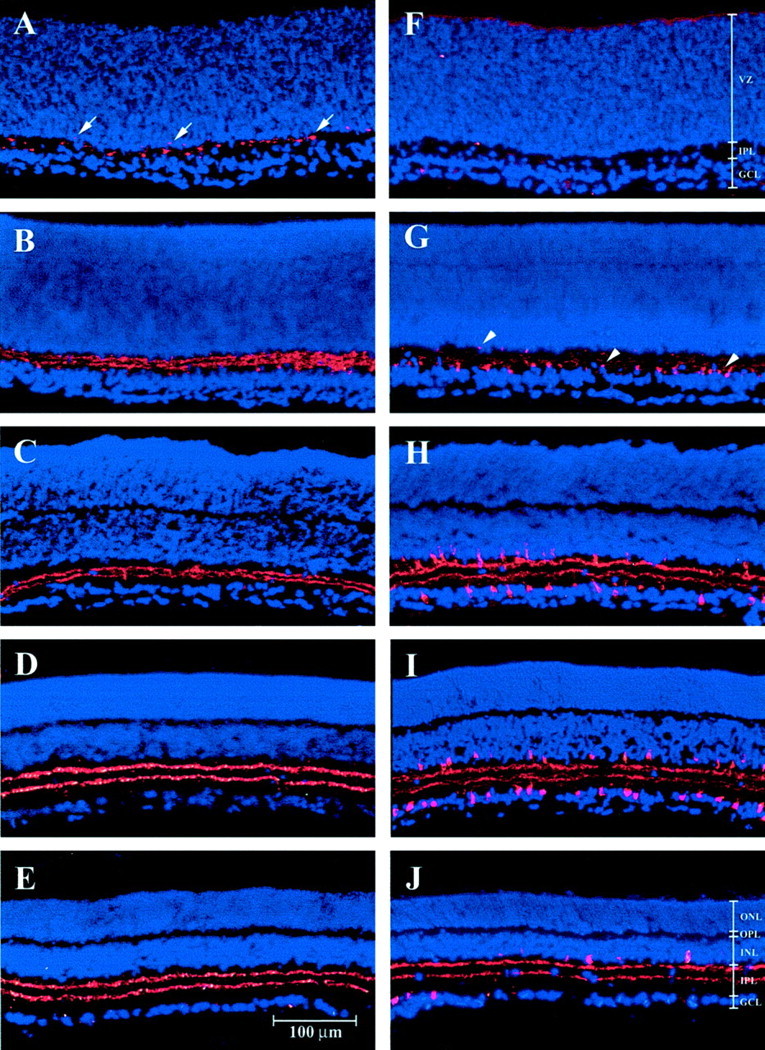
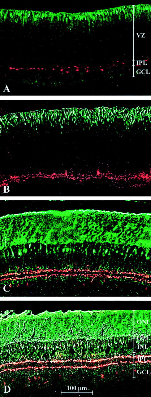
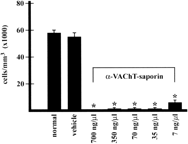
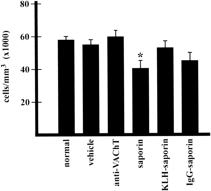
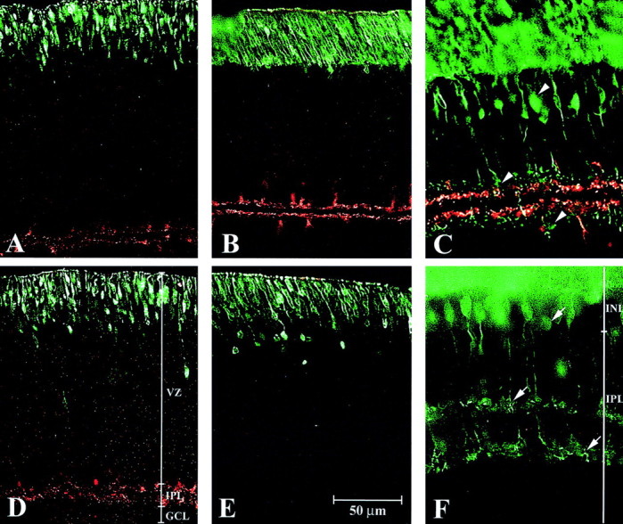
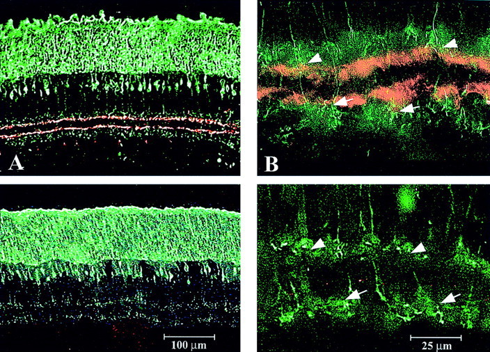
Cone bipolar cells are the first retinal neurons that respond in a differential manner to light onset and offset. In the mature retina, the terminal arbors of On and Off cone bipolar cells terminate in different sublaminas of the inner plexiform layer (IPL) where they form synapses with the dendrites of On and Off retinal ganglion cells and with the stratified processes of cholinergic amacrine cells. Here we first show that cholinergic processes within the On and Off sublaminas of the IPL are present early in development, being evident in the rat on the day of birth, approximately 10 d before the formation of segregated cone bipolar cell axons. This temporal sequence, as well as our previous finding that the segregation of On and Off cone bipolar cell inputs occurs in the absence of retinal ganglion cells, suggested that cholinergic amacrine cells could provide a scaffold for the subsequent in-growth of bipolar cell axons. To test this hypothesis directly, a new cholinergic cell immunotoxin was constructed by conjugating saporin, the ribosome-inactivating protein toxin, to an antibody against the vesicular acetylcholine transporter. A single intraocular injection of the immunotoxin caused a rapid, complete, and selective loss of cholinergic amacrine cells from the developing rat retina. On and Off cone bipolar cells were visualized using an antibody against recoverin, the calcium-binding protein that labels the soma and processes of these interneurons. After complete depletion of cholinergic amacrine cells, cone bipolar cell axon terminals still formed their two characteristic strata within the IPL. These findings demonstrate that the presence of cholinergic amacrine cells is not required for the segregation of recoverin-positive On and Off cone bipolar cell projections.


Figures

Fig. 1.
Fig. 1.
Cholinergic amacrine cells in the developing rat retina. The images are vertical sections of retinas with the inner, or corneal, side at the bottom of the section. VAChT antibody labeling is shown on the left, with ChAT labeling on the right (red). Sections are counterstained with DAPI nuclear stain (blue). Fromtop to bottom, the images show protein expression on the day of birth (P0) (A,F), P2 (B, G), P6 (C, H), P12 (D,I), and in the adult (E,J). The layers of the immature postnatal retina (F) include the ventricular zone (VZ), the inner plexiform layer (IPL), and the ganglion cell layer (GCL). In the mature retina (J), the layers are the outer nuclear, or photoreceptor, layer (ONL), the outer plexiform layer (OPL), the inner nuclear layer (INL), the IPL, and the GCL. Arrows show the earliest VAChT staining at P0 (A), and arrowheadsshow the earliest ChAT staining at P2 (G).
Fig. 2.
Fig. 2.
Cholinergic amacrine cells stratify before in-growth of cone bipolar cells. Shown are vertical sections of normal retinas stained with VAChT for cholinergic amacrine cells (red) and recoverin for cone bipolar cells (green). From top tobottom, the images show marker expression on P0 (A), P6 (B), P12 (C), and P20 (D). The layers of the immature (A) and mature retinas (D) are the same as in Figure 1. Note that the appearance and stratification of cholinergic amacrine cells precede the migration of cone bipolar cell bodies from the ventricular zone and the subsequent extension of their axonal processes into the IPL.
Fig. 3.
Fig. 3.
Determination of lowest effective dose of the immunotoxin. The vertical axis is the number of cholinergic amacrine cells per cubic millimeter of retina from morphometric analysis of ChAT immunoreactivity. The horizontal axis is the range of immunotoxin concentrations and vehicle injected in 2 μl total volume at P1. Counts were made at P12–P20. Values are ± SEM. Significance = p < 0.05. *p < 0.001.
Fig. 4.
Fig. 4.
Elimination of cholinergic neurons in the developing retina and adult brain. Shown are vertical sections immunostained for ChAT (red) against a DAPI background (blue). Retinas were injected at P1 with vehicle (left) or immunotoxin (right).A and B are P2 retinas; Cand D are P6 retinas. Arrows show detectible changes in ChAT immunoreactivity 24 hr after immunotoxin injection, whereas arrowheads show complete loss of cholinergic amacrine cells by P6.
Fig. 5.
Fig. 5.
Control series for immunotoxin structure and function. The number of cholinergic amacrine cells labeled with ChAT (per cubic millimeter) from a series of controls for immunotoxin structure and function. Values are ± SEM. Significance =p < 0.05. Note that only free saporin affected cholinergic amacrine cell number, reducing the number of cells ∼30% (*p < 0.001).
Fig. 6.
Fig. 6.
Cone bipolar cell in-growth proceeds normally.AF are P1-injected rat retinas double labeled with recoverin (green) and VAChT (red) killed at P2 (A, D), P6 (B, E), and P12 (C,F). The top images are vehicle-injected retinas, and the bottom images are toxin-injected retinas. Note the similarities between the staining pattern in the top series of panels and those in Figures 1 and 2, and that the stratification of cholinergic amacrine cells precedes the migration of cone bipolar cells. Note also that, even in the complete absence of VAChT immunoreactivity, cone bipolar cell somas migrate from the ventricular zone (E) and extend their axons to two distinct strata in the IPL (F), much as do cells in vehicle-treated retinas.
Fig. 7.
Fig. 7.
Axonal targeting of bipolar cells is tightly controlled. Shown are retinal cross sections with the GCL at the bottom, double immunostained with anti-recoverin for cone bipolar cells (green) and with anti-VAChT for cholinergic amacrine cells (red). Thetop panels are vehicle-injected retinas, and thebottom panels are toxin-injected retinas. The two low-magnification panels on the left show that the general morphology of the retina, particularly that of the cone bipolar cells, is unaffected by toxin treatment. The higher-magnification images on the right show that fine cone bipolar cell structures and axonal targeting are also primarily unaffected by the loss of cholinergic amacrine cells. Arrows show On bipolar terminals in the inner portion of the IPL, andarrowheads show Off bipolar terminals in the outer IPL.

Similar articles

Cited by

References

    1. Batchelor PE, Armstrong DM, Blaker SN, Gage FH. Nerve growth factor receptor and choline acetyltransferase colocalization in neurons within the rat forebrain: response to fimbria-fornix transection. J Comp Neurol. 1989;284:187–204. - PubMed
    1. Bisti S, Gargini C, Chalupa LM. Blockade of glutamate-mediated activity in the developing retina perturbs the functional segregation of ON and OFF pathways. J Neurosci. 1998;18:5019–5025. - PMC - PubMed
    1. Bodnarenko SR, Chalupa LM. Stratification of ON and OFF ganglion cell dendrites depends on glutamate-mediated afferent activity in the developing retina. Nature. 1993;364:144–146. - PubMed
    1. Bodnarenko SR, Jeyarasasingam G, Chalupa LM. Development and regulation of dendritic stratification in retinal ganglion cells by glutamate-mediated afferent activity. J Neurosci. 1995;15:7037–7045. - PMC - PubMed
    1. Brown SP, Masland RH. Costratification of a population of bipolar cells with the direction-selective circuitry of the rabbit retina. J Comp Neurol. 1999;408:97–106. - PubMed

Publication types

MeSH terms

LinkOut - more resources