. 2002 Mar 15;22(6):2299-312.
doi: 10.1523/JNEUROSCI.22-06-02299.2002.

Serotonin release evoked by tail nerve stimulation in the CNS of aplysia: characterization and relationship to heterosynaptic plasticity

Affiliations

Serotonin release evoked by tail nerve stimulation in the CNS of aplysia: characterization and relationship to heterosynaptic plasticity

Stephane Marinesco et al. J Neurosci. .

Abstract

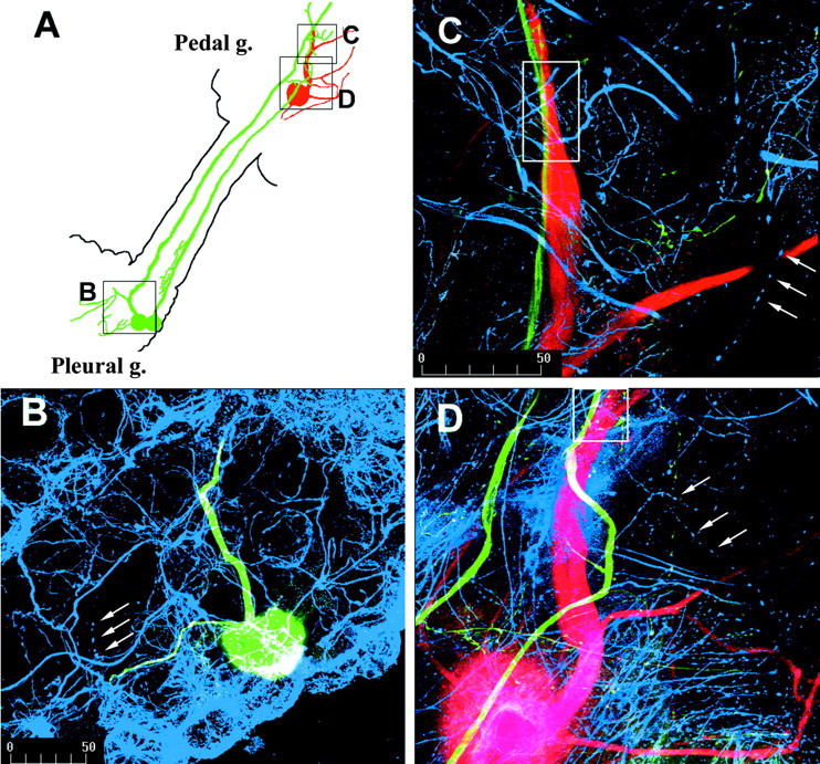
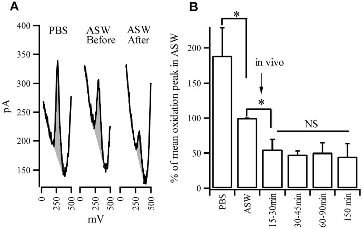
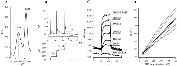
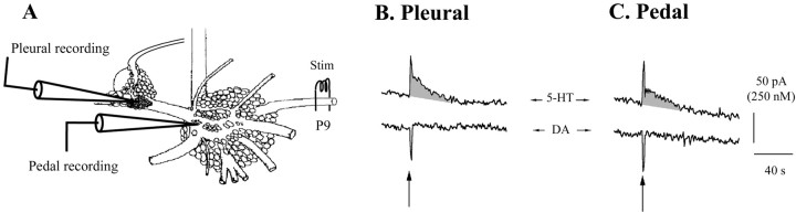
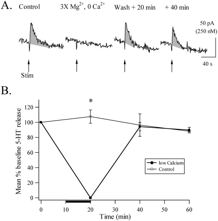
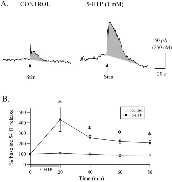
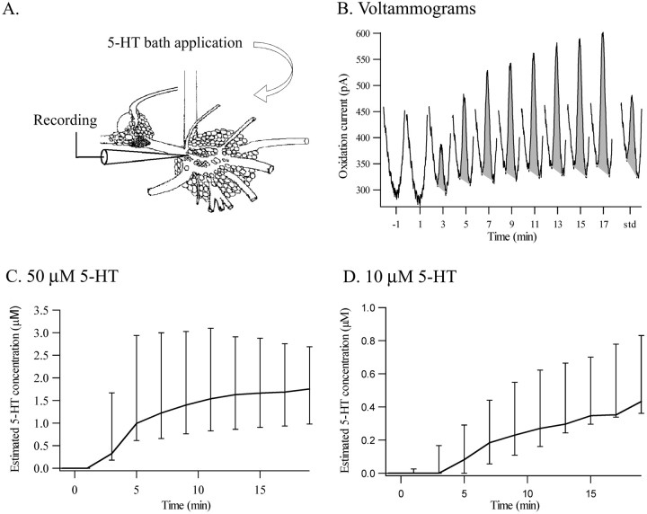
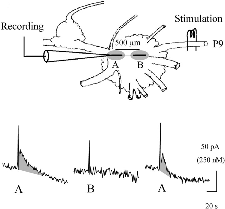
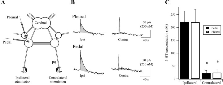
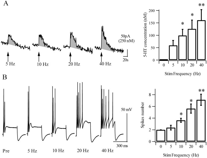
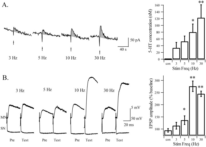
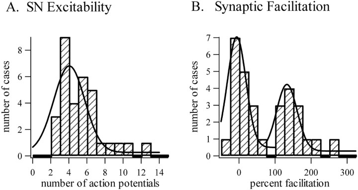
Considerable experimental evidence suggests that serotonin (5-HT) at sensory neuron-->motor neuron (SN-->MN) synapses, as well as other neuronal sites, contributes importantly to simple forms of learning such as sensitization and classical conditioning in Aplysia. However, the actual release of 5-HT in the CNS induced by sensitizing stimuli such as tail shock has not been directly demonstrated. In this study, we addressed this question by (1) immunohistochemically labeling central 5-HT processes and (2) directly measuring with chronoamperometry the release of 5-HT induced by pedal tail nerve (P9) shock onto tail SNs in the pleural ganglion and their synapses onto tail MNs in the pedal ganglion. We found that numerous 5-HT-immunoreactive fibers surround both the SN cell bodies in the pleural ganglion and SN axons in the pedal ganglion. Chronoamperometric detection of 5-HT performed with carbon fiber electrodes implanted in the vicinity of tail SN somata and synapses revealed an electrochemical 5-HT signal lasting approximately 40 sec after a brief shock of P9. 5-HT release was restricted to discrete subregions (modulatory fields) of the CNS, including the vicinity of tail SN soma and synapses ipsilateral to the stimulation. Increasing P9 shock frequency augmented the amplitude of the 5-HT signal and, in parallel, increased SN excitability and SN synaptic transmission onto tail MNs. However, the relationship between the amount of 5-HT release and the two forms of SN plasticity was not uniform: SN excitability increased in a graded manner with increased 5-HT release, whereas synaptic facilitation exhibited a highly nonlinear relationship. The development of chronoamperometric techniques in Aplysia now paves the way for a more complete understanding of the contribution of the serotonergic modulatory pathway to memory processing in this system.


Figures

Fig. 1.
Fig. 1.
Immunohistochemical localization of serotonergic fibers in close proximity to tail SNs. A, Schematic diagram of the preparation. Two SNs (green) in the pleural ganglion (g.) were identified in electrophysiological experiments as monosynaptically connected to a tail MN (red) in the pedal ganglion. Serotonergic fibers (blue) envelop the SN cell bodies in the pleural ganglion. Only one stained soma is apparent in the confocal plane. 5-HT-immunoreactive neurites overlaying the stained SN cell body appearwhite (B). Serotonergic fibers also travel into the pedal ganglion (C, D), where the SNs are known to synapse onto the MNs. Arrowheads show serotonergic varicosities. White boxes show examples of regions where 5-HT, SN, and MN processes are in close proximity. Scale is the same in B and D.
Fig. 2.
Fig. 2.
Effects of ASW and nervous tissue on electrode sensitivity for 5-HT. A, Examples of DNPV recordings obtained in a solution of 200 nm 5-HT diluted in PBS and ASW before implantation in the nervous tissue and after implantation. Oxidation peaks are highlighted by shading. Sensitivity is assessed by the height of the oxidation peak. It is greater in PBS than in ASW, and implantation of the electrode in the nervous tissue further decreases 5-HT sensitivity. B, Summary experiments of all DNPV recordings. Histograms represent the height of 5-HT oxidation peaks before and after a 15–150 min implantation in ganglia. The loss of sensitivity after implantation in the ganglion is similar after 15, 30, 60, and 150 min spent in the ganglion, suggesting stable reliable 5-HT detection in this time range.NS, Not significant. ∗p < 0.05.
Fig. 3.
Fig. 3.
Chronoamperometry allows rapid and selective detection of 5-HT. A, DA and 5-HT are oxidized, respectively, at 170 and 340 mV versus Ag/AgCl. DNPV recordings were obtained in a solution containing 200 nm 5-HT and 1 μm DA diluted in ASW. B, Chronoamperometry measurements were made with four pulses at 80, 230, 250, and 400 mV. The difference in the current at the end of the first two pulses represents substances oxidizing in the 80–230 mV range (DA), whereas the difference measured at the end of the last two pulses (δI) reflects oxidation of compounds in the 250–400 mV range (5-HT).C, Chronoamperometric recordings obtained in a flow-injection chamber where 5-HT was injected at various concentrations for 1 min. The corresponding electrodes had already been implanted in a ganglion. The bottom recording was performed on injection of DA (500 nm). D, The difference δI is linear with 5-HT concentration through the relation [5-HT] (nanomolar) ∼ 5.2 × δI (picoamperes). The light lines reflect individual electrodes; the bold line reflects the best linear fit to the responses of all electrodes.
Fig. 4.
Fig. 4.
Typical chronoamperometric recordings obtained in the CNS. The CFEs were implanted in the pleural ganglion, under the SN cell bodies and in the pedal ganglion, under the MN cell bodies (A, schematic diagram). The recorded oxidation current (δI) is increased after tail nerve shock in the 5-HT range (top pair of traces) but not in the DA range (bottom pair of traces). 5-HT signals were similar in the pleural (B) and the pedal (C) ganglia. The area under the 5-HT signal is highlighted by shading in this and subsequent figures. A 2 sec stimulation artifact was generated during tail nerve stimulation.
Fig. 5.
Fig. 5.
The electrochemical signal evoked by tail nerve stimulation relies on chemical synaptic transmission. The 5-HT signal is reversibly blocked by perfusion of 3× Mg2+, 0 Ca2+ ASW (see Materials and Methods).A, Individual traces obtained before, during, and after Ca2+ omission. B, Summary data obtained with six experiments. Low-Ca2+ treatment resulted in a complete (0%) block of the signal in all preparations. The amplitude of the signal was restored to its control level after 20 min in normal ASW. Error bars indicate SEM. ∗p < 0.05.
Fig. 6.
Fig. 6.
The 5-HT precursor 5-HTP increases the electrochemical signal evoked by a tail nerve shock. A, Typical recordings before and after 5-HTP application.B, Summary data showing that the effect of 5-HTP lasts at least 1 hr after washout (n = 8). * indicates significant difference from control recordings (p < 0.05). Error bars indicate SEM. Data are normalized to the amplitude of the first 5-HT signal at time0. Stim, Stimulation.
Fig. 7.
Fig. 7.
Exogenously applied 5-HT reaches physiological concentrations in the tail SN→MN synaptic neuropil. A, Schematic representation of the preparation. B, Representative voltammograms measured in the pedal SN→MN synaptic neuropil during perfusion of 50 μm 5-HT into the bath. Oxidation peaks at the 5-HT oxidation potential (340 mV) areshaded. At the end of the experiment, the electrode was removed from the ganglion and tested in a standard solution of 500 nm 5-HT (std) to estimate the concentrations that had reached the neuropil during 5-HT perfusion. C, Summary graph of estimated 5-HT concentrations in the SN→MN synaptic region during perfusion of 50 μm 5-HT (data are medians ± interquartile range; n = 8).D, Summary graph for 10 μm 5-HT (data as in C). The median concentration after 5 min of 5-HT is 80 and 990 nm (for 10 and 50 μm,respectively), which is close to the estimated 5-HT release evoked by tail nerve shock (see Results).
Fig. 8.
Fig. 8.
The 5-HT signal in the pedal ganglion is localized to a discrete subregion of the neuropil. Top, The carbon fiber electrode was inserted in the pedal ganglion, underneath the MN cell bodies for the first recording (region A), and moved 500 μm more caudal for the second recording (region B). Bottom, The 5-HT signal was maximal in the MN area, undetectable 500 μm more caudal, and restored when the electrode was moved back to region A.
Fig. 9.
Fig. 9.
The 5-HT signal is lateralized to the ipsilateral side of the stimulation. A, Schematic view of the ring ganglia, ventral side up, with both tail nerves connected to a stimulating electrode. B, Chronoamperometric recordings were performed in the pleural and pedal ganglia. The 5-HT signal was maximal after ipsilateral (Ipsi) tail nerve stimulation and was barely detectable after a contralateral (Contra) shock. C, Summary of six different preparations. The 5-HT signal evoked by contralateral stimulation was ∼10% of the one induced by ipsilateral stimulation. Error bars indicate SEM. * indicates significant difference between ipsilateral and contralateral stimulation,p < 0.05.
Fig. 10.
Fig. 10.
Varying the frequency of tail nerve stimulation modulates the amplitude of 5-HT release and its ability to increase SN excitability. A, Representative 5-HT responses evoked by 2 sec tail nerve shocks with frequencies ranging from 5 to 40 Hz applied at 30 min intervals. Right panel, Summary of chronoamperometry data. The evoked 5-HT signal progressively increases with shock frequency, ranging from 50 nm (5 Hz) to 170 nm (40 Hz) (n = 6). B, Example of SN excitability after 5, 10, 20, and 40 Hz stimulation.Right panel, Excitability also increases progressively with shock frequency (n = 8). Error bars indicate SEM. * and ** indicate significant difference from control and 5 Hz conditions, respectively (see Results). Stim, Stimulation.
Fig. 11.
Fig. 11.
Effects of tail nerve shock on facilitation of SN→MN synapses. A, Examples of 5-HT signals evoked by 2 sec shock frequencies ranging from 3 to 30 Hz applied at 30 min intervals.Right panel, Mean 5-HT concentrations evoked by tail nerve stimulation (Stim) at the same frequencies (Freq) (n = 6). As before (Fig. 9), the signal progressively increases with stronger stimulation.B, Examples of the effects of tail nerve shock on an SN→MN synapse. STF is induced at 30 and 10 but not at 3 Hz; a small but significant facilitation appears at 5 Hz. A discontinuity in the induction of STF appears between 5 and 10 Hz, corresponding to ∼60 nm 5-HT. * indicates significant difference from no shock; ** indicates significant difference with combined control (con) 3–5 Hz conditions (see Results). Pre, Before P9 shock.
Fig. 12.
Fig. 12.
Frequency histogram analysis of the effects of tail nerve stimulation on SN plasticity. Individual experiments aimed at assessing increases in SN excitability or facilitation of SN→MN synapses are plotted in A and B, respectively. Whereas excitability increases continuously with shock frequency, synaptic facilitation analysis shows two well separated groups of data: one centered around 0 (nonfacilitated synapses) and the other at approximately +150% (facilitated synapses).

Similar articles

Cited by

References

    1. Abrams TW, Castellucci VF, Camardo JS, Kandel ER, Lloyd PE. Two endogenous neuropeptides modulate the gill and siphon withdrawal reflex in Aplysia by presynaptic facilitation involving cAMP-dependent closure of a serotonin-sensitive potassium channel. Proc Natl Acad Sci USA. 1984;81:7956–7960. - PMC - PubMed
    1. Alberini C, Ghirardi M, Metz R, Kandel E. C/EBP is an immediate-early gene required for the consolidation of long-term facilitation in Aplysia. Cell. 1994;76:1099–1114. - PubMed
    1. Angers A, Storozhuk MV, Duchaine T, Castellucci VF, DesGroseillers L. Cloning and functional expression of an Aplysia 5-HT receptor negatively coupled to adenylate cyclase. J Neurosci. 1998;18:5586–5593. - PMC - PubMed
    1. Barbas D, Zappulla J, Angers S, Bouvier M, Castellucci V, DesGroseillers L. Functional characterization of a novel serotonin receptor (5-HTap2) expressed in the CNS of Aplysia californica. J Neurochem. 2002;80:335–345. - PubMed
    1. Brabham DG, Fernandez RI, Levenson J, Eichberg J, Byrne JH, Eskin A. Diurnal regulation of serotonin synthesis and expression of sensitization in Aplysia. Soc Neurosci Abstr. 2000;26:1525.

Publication types

MeSH terms