. 2002 Mar 15;22(6):2383-90.
doi: 10.1523/JNEUROSCI.22-06-02383.2002.

Elevated fusiform cell activity in the dorsal cochlear nucleus of chinchillas with psychophysical evidence of tinnitus

Affiliations

Elevated fusiform cell activity in the dorsal cochlear nucleus of chinchillas with psychophysical evidence of tinnitus

T J Brozoski et al. J Neurosci. .

Abstract

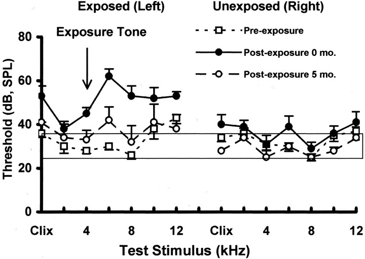
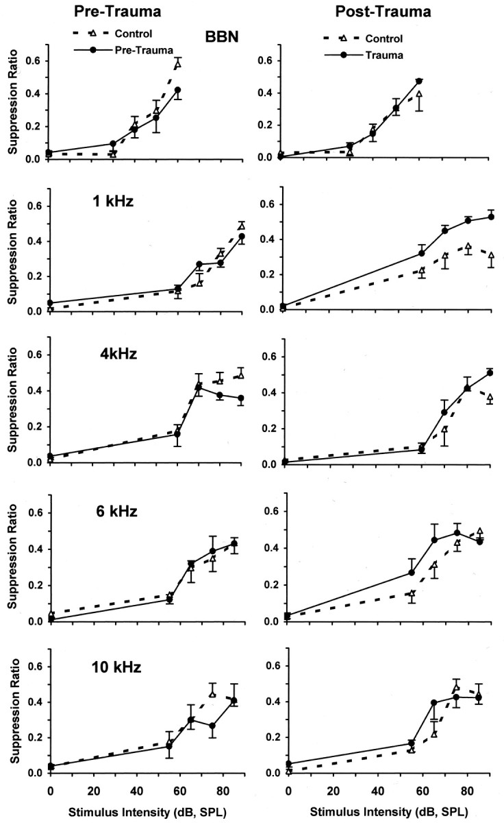
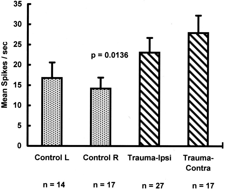
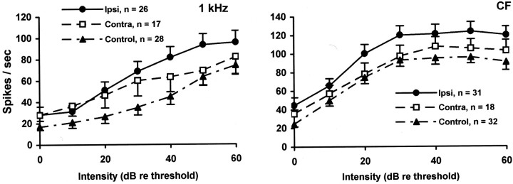
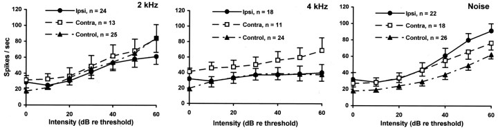
Chinchillas with psychophysical evidence of chronic tinnitus were shown to have significantly elevated spontaneous activity and stimulus-evoked responses in putative fusiform cells of the dorsal cochlear nuclei (DCN). Chinchillas were psychophysically trained and tested before and after exposure to a traumatic unilateral 80 dB (sound pressure level) 4 kHz tone. Before exposure, two groups were matched in terms of auditory discrimination performance (noise, and 1, 4, 6, and 10 kHz tones). After exposure, a single psychophysical difference emerged between groups. The exposed group displayed enhanced discrimination of 1 kHz tones (p = 0.00027). Postexposure discrimination of other stimuli was unaffected. It was hypothesized that exposed animals experienced a chronic subjective tone (i.e., tinnitus), resulting from their trauma, and that features of this subjective tone were similar enough to 1 kHz to affect discrimination of 1 kHz objective signals. After psychophysical testing, single-unit recordings were obtained from each animal's DCN fusiform cell layer. Putative fusiform cells of exposed animals showed significantly (p = 0.0136) elevated spontaneous activity, compared with cells of unexposed animals. Putative fusiform cells of exposed animals showed a greater stimulus-evoked response to tones at 1 kHz (p = 0.0000006) and at characteristic-frequency (p = 0.0000009). This increased activity was more pronounced on the exposed side. No increase in stimulus-evoked responses was observed to other frequencies or noise. These parallel psychophysical and electrophysiological results are consistent with the hypothesis that chronic tonal tinnitus is associated with, and may result from, trauma-induced elevation of activity of DCN fusiform cells.


Figures

Fig. 1.
Fig. 1.
Auditory thresholds before and after trauma. Thresholds were measured using acoustically evoked brainstem potentials, recorded immediately after stimulus onset. Shown are the mean threshold functions (error bars indicate 1 SEM) for trauma-exposed subjects (n = 4). The normative range was determined by the average, ± 1 SEM, of four unexposed control animals. The trauma stimulus is indicated by the arrow. Immediately after exposure there was a significant ipsilateral threshold elevation (left panel), compared with pre-exposure levels, for tones from 4 to 6 kHz (p = 0.005–0.007) and for clicks (p = 0.01) in the exposed ear (ipsilateral). There was no significant threshold elevation for 1 kHz tones immediately after trauma (p = 0.08), nor were there significant threshold elevations 5 months after trauma for any stimulus (p = 0.157–0.719).
Fig. 2.
Fig. 2.
Representative PSTH and RIFs recorded from putative fusiform cells in the chinchilla DCN. Stimuli were presented to the ipsilateral ear. Top panel, RIFs of a unit recorded from the ipsilateral DCN of a trauma-exposed subject and a unit recorded from the DCN of an unexposed control subject.Bottom panel, Poststimulus histogram of a “buildup” unit from the ipsilateral DCN of a trauma-exposed subject. These profiles are similar to those reported by others for DCN fusiform units (Young, 1980).
Fig. 3.
Fig. 3.
Pre-trauma versus post-trauma psychophysical discrimination functions (error bars indicate 1 SEM). Pre-trauma (left panels) and post-trauma (right panels) psychophysical discrimination functions for control (no trauma) and trauma-exposed subjects (n = 4/group) are shown. Test stimulus values are indicated in each panel. Suppression ratios reflect the discrimination performance of subjects, with a value of 0 indicating discrimination of 0 dB, and a value of 0.5 indicating discrimination of sound comparable to that of 60 dB broadband noise. The only significant difference between control (dashed lines) and trauma-exposed (solid lines) groups was for 1 kHz tones after trauma (see Table 1 for significance levels).
Fig. 4.
Fig. 4.
DCN fusiform cell spontaneous activity. Spontaneous activity of DCN fusiform cells in control and trauma-exposed chinchillas (n = 4 animals/group). Spontaneous activity was measured in single units as cumulative spikes over 40 sec epochs. Spontaneous activity is indicated as mean spikes per second (error bars indicate 1 SEM). Control data are presented for cells from the left and right DCN. Trauma data are presented for cells from the ipsilateral (left/exposed ear) and contralateral (right/unexposed ear) DCN. The number of units in each classification is shown below the x-axis. There was a significant difference in spontaneous activity between trauma fusiform cells (Trauma-Ipsi and Trauma-Contra) and control fusiform cells (Control L and Control R), with no significant hemilateral differences.
Fig. 5.
Fig. 5.
RIFs for 1 kHz (left panel) and CF (right panel) tones. RIFs are shown for putative fusiform cells from the ipsilateral (Ipsi) and contralateral (Contra) DCN of trauma-exposed animals, as well as controls (left and rightcombined; error bars indicate 1 SEM). The 1 kHz RIF of ipsilateral cells and contra cells was significantly elevated above that of controls, with the ipsilateral effect much more pronounced than the contralateral effect. The CF RIF of ipsilateral cells was significantly elevated above that of control cells as well as that of contralateral cells. The CF RIF of contra cells were not significantly different than that of controls. The number of cells contributing to each function is shown in the key. These electrophysiological results parallel the psychophysical results depicted in Figure 3.
Fig. 6.
Fig. 6.
RIFs for stimuli other than 1 kHz tones. RIFs are shown for putative fusiform cells from the ipsilateral (Ipsi) and contralateral (Contra) DCN of trauma-exposed animals, as well as controls (left andright combined; error bars indicate 1 SEM). There were no significant differences between the ipsilateral, contralateral, and control functions. The number of cells contributing to each function is shown in the key.

References

    1. Alibardi L. Cytology of large neurons in the guinea pig dorsal cochlear nucleus contacting the inferior colliculus. Eur J Histochem. 2000;44:365–375. - PubMed
    1. Anari M, Axelsson A, Eliasson A, Magnusson L. Hypersensitivity to sound–questionnaire data, audiometry and classification. Scand Audiol. 1999;28:219–230. - PubMed
    1. Atherley GRC, Hempstock TI, Noble WG. Study of tinnitus induced temporarily by noise. J Acoust Soc Am. 1968;44:1503–1506. - PubMed
    1. Backoff PM, Palombi PS, Caspary DM. Glycinergic and GABAergic inputs affect short-term suppression in the cochlear nucleus. Hear Res. 1997;110:155–163. - PubMed
    1. Bauer CA, Brozoski TJ. Assessing tinnitus and prospective tinnitus therapeutics using a psychophysical animal model. J Assoc Res Otolaryngol. 2001;2:54–64. - PMC - PubMed

Publication types