. 2002 Apr;3(4):341-8.
doi: 10.1093/embo-reports/kvf069. Epub 2002 Mar 15.

Plk1 promotes nuclear translocation of human Cdc25C during prophase

Affiliations

Plk1 promotes nuclear translocation of human Cdc25C during prophase

Fumiko Toyoshima-Morimoto et al. EMBO Rep. 2002 Apr.

Abstract

The nuclear accumulation of active M-phase promoting factor (MPF) during prophase is thought to be essential for coordinating M-phase events in vertebrate cells. The protein phosphatase Cdc25C, an activator of MPF, enters the nucleus to keep MPF active in the nucleus during prophase. However, the molecular mechanisms that control nuclear translocation of Cdc25C during prophase are unknown. We show that phosphorylation of a serine residue (Ser198) in a nuclear export signal sequence of human Cdc25C occurs during prophase and promotes nuclear localization of Cdc25C. We also show that Polo-like kinase 1 (Plk1) is responsible for this phosphorylation and that constitutively active Plk1 promotes nuclear localization of Cdc25C. Remarkably, a mutant Cdc25C in which Ser198 is replaced by alanine remains in the cytoplasm when wild-type Cdc25C accumulates in the nucleus during prophase. These results suggest that Plk1 phosphorylates Cdc25C on Ser198 and regulates nuclear translocation of Cdc25C during prophase.


Figures

None
Fig. 1. Serine phosphorylation in the NES sequence of human Cdc25C causes its nuclear localization. Indicated forms of HA-Cdc25C were expressed in HeLa cells. More than 200 cells were examined in each condition, and representative images are shown in the upper panels and DAPI staining shown below. Cells were classified into three categories in terms of location of the transfected HA-tagged Cdc25C, and the percentages of each categories are shown in the bottom graph, where N>C shows staining in the nucleus (N) stronger than that in the cytoplasm (C), N=C shows N equal to C, and N<C shows C stronger than N.
None
Fig. 2. Plk1 phosphorylates Ser198 of Cdc25C. (A) NES sequences of cyclin B1 (human) and Cdc25C (human). Important hydrophobic residues in NES are boxed. (B) The kinase activities of recombinant His-tagged Plk1, which was expressed in and purified from bacteria, and HA-tagged Plk1, which was expressed in COS cells and purified by immunoprecipitation with anti-HA, were measured using indicated forms of GST–Cdc25C (173–206). (C) A synthetic peptide corresponding to residues 188–201 (EEISDELMEFSLKD) of human Cdc25C (NES) or a synthetic phosphopeptide [EEISDELMEF-phosphoS198-LKD (phosphoS198-NES)] was conjugated to ovalbumin (OVA). The kinase activities of His-tagged Plk1 toward OVA, OVA-NES and OVA-P-NES are shown. (D) Bacterially produced GST–WT-Cdc25C (full length) and GST–S198A-Cdc25C (full length) were pre-incubated with or without His-Plk1 in the presence of ATP, subjected to SDS–PAGE, transfered to a nylon membrane and then blotted with anti-phosphoS198 Cdc25C antibody (top row). The bottom row shows CBB staining of GST–Cdc25C (full length) on the membrane.
None
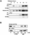
Fig. 3. Plk1 phosphorylates Cdc25C on Ser198 during G2–M phase in HeLa cells. (A) HeLa cells were arrested at the G1–S boundary by a double thymidine block, and cell extracts were prepared at the indicated times after release. The kinase activities in the extracts toward GST–WT-Cdc25C (173–206), S198A-Cdc25C (173–206) and histone H1 are shown in the first, second and third rows, respectively. Endogenous Plk1 was immunoprecipitated from the extracts with anti-Plk1 antibody. The kinase activities of the immunoprecipitates toward GST–WT-Cdc25C (173–206) and S198A-Cdc25C (173–206) are shown in the fourth and fifth rows, respectively. (B) Endogenous Plk1 was immunoprecipitated from the synchronized M-phase extracts with increasing amounts (0, 2.5 and 7.5 µg) of anti-Plk1 antibody or non-immunized rabbit IgG, and the kinase activity remaining in the supernatant was measured using GST–WT-Cdc25C (173–206) as a substrate (top row). The supernatants were analyzed by SDS–PAGE followed by immunoblotting with anti-Plk1 antibody (bottom row).
None
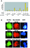
Fig. 4. Constitutively active Plk1 causes nuclear translocation of Cdc25C. (A) The indicated forms of Myc-tagged Cdc25C, HA-tagged Plk1 (vector, WT or SDTD) and/or untagged cyclin B1 were expressed in HeLa cells. Fifteen hours after transfection, cells were fixed, followed by staining with anti-Myc and anti-HA antibodies. More than 200 cells were examined in each condition, and the percentages of the cells in which Myc-tagged Cdc25C was accumulated in the nucleus are shown. (B) Typical images of Myc-tagged Cdc25C (middle column) co-expressed with HA-tagged Plk1 (left column) and untagged cyclin B1 are shown with DAPI staining (right column). When S198,216A-Cdc25C was expressed, part of this mutant Cdc25C localized to the nucleus. Expression of SDTD-Plk1 and cyclin B1 did not enhance at all the nuclear localization of this mutant Cdc25C (data not shown).
None
Fig. 5. Phosphorylation of Ser198 of Cdc25C occurs during G2–M phase in vivo and is necessary for its nuclear translocation. (A) Indicated amounts of OVA-NES and OVA-phosphoS198-NES were immunoblotted with the affinity-purified anti-phosphoSer198 Cdc25C antibody (α-phosphoS198). (B) Cell extracts were prepared from synchronized HeLa cells at 4 h (S phase) and 10 h (M phase) after release from a double thymidine block, analyzed by SDS–PAGE and subjected to immunoblotting with anti-Cdc25C antibody (left panel) or α-phosphoS198 (right panel). α-PhosphoS198 reacted with both a 55-kDa polypeptide in M-phase extracts (arrow), which corresponds to S198-phosphorylated Cdc25C, and an uncharacterized 60-kDa polypeptide (arrowhead). (C) S-phase and M-phase extracts were subjected to immunoprecipitation with α-phosphoS198. The extracts (bottom row) and the immunoprecipitates (top row) were immunoblotted with anti-Cdc25C antibody. (D) HeLa cells were synchronized by a double thymidine block, and cell extracts were prepared at the indicated times after release. The kinase activities in the extracts toward histone H1 are shown in the top graph. Endogenous Plk1 was immunoprecipitated from the extracts with anti-Plk1 antibody, and the kinase activities of the immunoprecipitates toward GST–WT-Cdc25C (173–206) are shown in the bottom graph. The extracts were analyzed by SDS–PAGE followed by immunoblotting with anti-Cdc25C antibody and α-phosphoS198 (bottom panels). (E) Myc-tagged WT-Cdc25C or S198A-Cdc25C was expressed in HeLa cells. The cells were then arrested in M phase by nocodazole treatment. The cell extracts were prepared and subjected to immunoprecipitaion with anti-Myc antibody. The immunoprecipitates were treated with or without protein phosphatase PP1 and subjected to immunoblotting with α-phosphoS198S and anti-Myc antibodies. (F) Myc-tagged WT-Cdc25C and HA-tagged S198A-Cdc25C were expressed in HeLa cells. Cells were fixed and stained with anti-Myc and anti-HA antibodies. A typical image is shown. This image was detected in 13 out of 15 prophase cells.

Similar articles

Cited by

References

    1. Blasina A., Van de Weyer, M., Laus, M.C., Luyten, W.H., Parker, A.E. and McGowan, C.H. (1999) A human homologue of the checkpoint kinase Cds1 directly inhibits Cdc25 phosphatase. Curr. Biol., 9, 1–10. - PubMed
    1. Chen M.S., Hurov, J., White, L.S., Woodford-Thomas, T. and Piwnica-Worms, H. (2001) Absence of apparent phenotype in mice lacking Cdc25C protein phosphatase. Mol. Cell. Biol., 12, 3853–3861. - PMC - PubMed
    1. Coleman T.R. and Dunphy, W.G. (1994) Cdc2 regulatory factors. Curr. Opin. Cell Biol., 6, 877–882. - PubMed
    1. Dalal S.N., Schweitzer, C.M., Gan, J. and DeCaprio, J.A. (1999) Cytoplasmic localization of human cdc25C during interphase requires an intact 14-3-3 binding site. Mol. Cell. Biol., 19, 4465–4479. - PMC - PubMed
    1. Furnari B., Rhind, N. and Russell, P. (1997) Cdc25 mitotic inducer targeted by chk1 DNA damage checkpoint kinase. Science, 277, 1450–1451. - PubMed

Publication types