. 2002 Mar 15;539(Pt 3):681-91.
doi: 10.1113/jphysiol.2001.013246.

Alpha(1H) mRNA in single skeletal muscle fibres accounts for T-type calcium current transient expression during fetal development in mice

Affiliations

Alpha(1H) mRNA in single skeletal muscle fibres accounts for T-type calcium current transient expression during fetal development in mice

Christine Berthier et al. J Physiol. .

Abstract

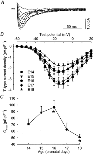
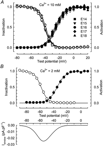
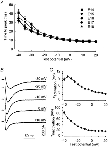
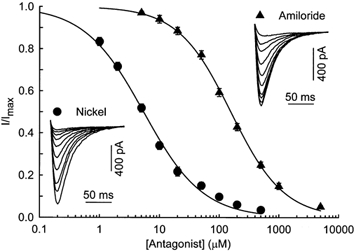
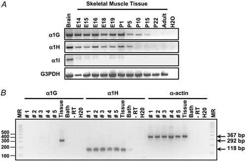
Calcium channels are essential for excitation-contraction coupling and muscle development. At the end of fetal life, two types of Ca(2+) currents can be recorded in muscle cells. Whereas L-type Ca(2+) channels have been extensively studied, T-type channels have been poorly characterized in skeletal muscle. We describe here the functional and molecular properties of T-type calcium channels in developing mouse skeletal muscle. The T-type current density increased transiently during prenatal myogenesis with a maximum at embryonic day E16 followed by a drastic decrease until birth. This current showed similar electrophysiological and pharmacological properties at all examined stages. It displayed a wide window current centred at about -35 and -55 mV in 10 and 2 mM external Ca(2+), respectively. Activation and inactivation kinetics were fast (3 and 16 ms, respectively). The current was inhibited by nickel and amiloride with an IC(50) of 5.4 and 156 microM, respectively, values similar to those described for cloned T-type alpha(1H) channels. Whole muscle tissue RT-PCR analysis revealed mRNAs corresponding to alpha(1H) and alpha(1G) subunits in the fetus but not in the adult. However, single-fibre RT-PCR demonstrated that only alpha(1H) mRNA was present in prenatal fibres, suggesting that the alpha(1G) transcript present in muscle tissue must be expressed by non-skeletal muscle cells. Altogether, these results demonstrate that the alpha(1H) subunit generates functional T-type calcium channels in developing skeletal muscle fibres and suggest that these channels are involved in the early stages of muscle differentiation.


Figures

Figure 1
Figure 1. Voltage dependence of T-type Ca2+ current density during prenatal myogenesis
A, superimposition of difference traces, in response to test pulses to −45, −40, −35, −30, −25, −15, −5, 5 and 15 mV. The cell was isolated from a fetus at E16 and had a capacitance of 187 pF. B, voltage dependence of the average density of T-type Ca2+ current recorded in myofibres from 14- (n = 13), 15- (n = 10), 16- (n = 15), 17- (n = 8) and 18-day-old (n = 8) fetuses. The curves correspond to a fit to the mean data at each age using eqn (1) from Methods. C, average maximum macroscopic conductance calculated from the I-V curve as described in Methods plotted versus the age of the fetuses. Asterisks indicate two values which are significantly different (ANOVA test, P < 0.05).
Figure 2
Figure 2. Properties of the T-type Ca2+ current during prenatal myogenesis
A, voltage dependence of T-type Ca2+ current activation (filled symbols) and inactivation (open symbols) in myofibres from 14- (n = 13), 15- (n = 10), 16- (n = 15), 17- (n = 8) and 18-day-old (n = 8) fetuses in the presence of 10 mm external Ca2+. The data for the activation and inactivation in each cell were obtained as described in Methods and then averaged on this graph. Continuous lines correspond to a Boltzmann fit to the population average. B, top, same curves as in A but in the presence of 2 mm external Ca2+. The data were obtained from 9 cells at E16. Parameters of the fits are V1/2,ac=−34.8 mV, kac= 6.36 mV, V1/2,inac=−58.6 mV and kinac=−5.00 mV. Bottom, putative window current obtained by multiplying the predicted peak current-voltage curve by the predicted steady-state inactivation curve. The average window current reached a maximum of 0.025 pA pF−1 at a potential of −54.8 mV.
Figure 3
Figure 3. Kinetics of the T-type Ca2+ current
A, voltage dependence of the time to peak in myofibres from 14- (n = 13), 15- (n = 10), 16- (n = 15), 17- (n = 8) and 18-day-old (n = 8) fetuses. B, T-type Ca2+ current evoked in response to test depolarizations from a holding potential of −80 mV to the indicated potentials. The continuous lines correspond to a fit to the experimental data using eqn (3) described in Methods. Capacitance of the cell isolated from a 16-day-old fetus was 111.5 pF. C, voltage dependence of the activation (top) and inactivation (bottom) time constants of the T-type Ca2+ current. Values are means ± s.e.m. of 20 cells at E16.
Figure 4
Figure 4. Time dependence of recovery from short-term inactivation
Average plots (from 12 cells at E16) of the relative peak current elicited by the test pulse as a function of the interpulse duration. The relationship was fitted by a double exponential with a time constant of 224 ms for 63 % of the current and 5.16 s for the other 37 % of the current. Inset, superimposed current traces from the beginning of a typical experiment showing the increase of the current induced by the test pulse when the recovery interval grew longer. The time dependence of recovery was studied using a paired pulse protocol (see below the traces) applied every 60 s and where inactivation was induced by a 250 ms depolarization to −20 mV. Capacitance of the cell isolated from a 16-day-old fetus was 302 pF.
Figure 5
Figure 5. Effect of nickel ions and amiloride on T-type Ca2+ current
Dose-response curve for the block of T-type Ca2+ current by the antagonist. Data represent the average (± s.e.m.) responses from 9 and 7 cells at E16, for NiCl2 and amiloride, respectively. The smooth curves represent the fit of the Hill equation to the data with IC50= 5.4 and 156 μm and nH= 0.88 and 0.98 for NiCl2 and amiloride, respectively. Insets, typical responses to increasing concentrations of NiCl2 and amiloride. Test pulses to −20 mV from a holding potential of −80 mV were delivered every 20 s. Traces shown here are control traces and current in presence of 1, 2, 5, 10, 20, 50, 100, 200 and 500 μm NiCl2 or 5, 10, 20, 50, 100, 200, 500, 1000 and 5000 μm amiloride. Capacitance of the cells isolated from 16-day-old fetuses was 152 and 109 pF, respectively.
Figure 6
Figure 6. Expression of the mRNAs corresponding to the α1G, α1H and α1I T-type Ca2+ channel subunits during skeletal muscle development
A, α1G, α1H and α1I transcripts were detected by RT-PCR in muscle tissue samples from various embryonic (E) and postnatal (P) stages and from adult mouse using specific forward and reverse primers as described in Methods. Brain sample was used as a positive control and water as a negative control. G3PDH amplification was performed on all samples as a positive control. B, single-fibre RT-PCR analysis on isolated fibres from 18-day-old fetuses. Expression of α1G, α1H and skeletal muscle α-actin transcripts was analysed on five different fibres (lanes numbered 1 to 5) and on 50 × 10−12 g of RNA from whole skeletal muscle tissue (lane ‘Tissue’). Note that PCR products corresponding to α1H (118 bp) and α-actin (367 bp) transcripts are present in all analysed fibres and in whole muscle tissue whereas the PCR product corresponding to the α1G subunit transcript (292 bp) is only detected in muscle tissue. Negative controls are the corresponding bath solution, a single-fibre sample without reverse transcriptase and water. The size of the different fragments in the molecular ruler (MR, 100 bp PCR ladder) is shown on the left.

References

    1. Beam KG, Knudson CM. Calcium currents in embryonic and neonatal mammalian skeletal muscle. Journal of General Physiology. 1988a;91:781–798. - PMC - PubMed
    1. Beam KG, Knudson CM. Effect of postnatal development on calcium currents and slow charge movement in mammalian skeletal muscle. Journal of General Physiology. 1988b;91:799–815. - PMC - PubMed
    1. Bijlenga P, Liu J-H, Espinos E, Haenggli C-A, Fischer-Lougheed J, Bader CR, Bernheim L. T-type alpha1H Ca2+ channels are involved in Ca2+ signaling during terminal differentiation (fusion) of human myoblasts. Proceedings of the National Academy of Sciences of the USA. 2000;97:7627–7632. - PMC - PubMed
    1. Bossu J-L, Feltz A, Thomann JM. Depolarization elicits two distinct calcium currents in vertebrate sensory neurones. Pflügers Archiv. 1985;403:360–368. - PubMed
    1. Carbone E, Lux HD. A low voltage-activated, fully inactivating Ca channel in vertebrate sensory neurones. Nature. 1984;310:501–502. - PubMed

Publication types

LinkOut - more resources