Comparative Study
. 2002 Mar 15;539(Pt 3):725-33.
doi: 10.1113/jphysiol.2001.013407.

External anions and cations distinguish between AMPA and kainate receptor gating mechanisms

Affiliations
Comparative Study

External anions and cations distinguish between AMPA and kainate receptor gating mechanisms

Derek Bowie. J Physiol. .

Abstract

Experiments were designed to examine if ion-flow through alpha-amino-3-hydroxy-5-methyl-isoxazolepropionic acid (AMPA) or kainate receptors interferes with protein structures associated with the gating machinery. Gating was studied using ultra-fast drug perfusion of outside-out patches containing rat GluR-A or GluR6 subunits excised from transfected human embryonic kidney cells. Deactivation rates of GluR6 kainate receptors observed following brief L-glutamate (10 mM Glu, 1 ms) applications differed by two to threefold in high (405 mM symmetrical Na(+), tau(decay) = 2.7 ms at -100 mV) and low ionic strength (55 mM, tau(decay) = 1.1 ms) solutions. In comparison, GluR-A AMPA receptors were much less sensitive. Ion effects on GluR6 receptors did not reflect surface potential screening or ion-agonist competition at the agonist-binding site since deactivation rates were slower in high ionic strength solutions. Moreover, the apparent agonist affinity did not decrease with increasing ionic strength (e.g. 55 mM, EC(50) = 110 microM vs. 405 mM, EC(50) = 61 microM). GluR6 responses were strongly dependent on ions present on the external, but not the internal, side of the plasma membrane. Decay kinetics was regulated by the type of ion present suggesting that the chemical nature of the solution, not its ionic strength, governed channel behaviour. Both external anions and cations modulated the amplitude and decay kinetics of GluR6 responses in a concomitant manner. AMPA receptor responses recorded in identical ionic conditions did not exhibit this behaviour. These results identify a novel mechanism that distinguishes AMPA and kainate receptors. External ions regulate the gating machinery of kainate receptors through an allosteric mechanism that involves both anions and cations.


Figures
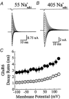
Figure 1
Figure 1. Ions affect the gating properties of kainate receptors
Membrane currents activated by brief (1 ms) applications of 10 mm l-Glu at various membrane potentials (−100 to + 125 mV, 15 mV increments) in 55 mm (A) and 405 mm (B) symmetrical NaCl. In each case, the top trace shows the junction current (1 ms) recorded with an open patch electrode at the end of the experiment. C, summary plot comparing the deactivation rates measured in low (55 mm Na+, ○, n = 11) and high (405 mm Na+, •, n = 3) ionic strength.
Figure 2
Figure 2. Ion effects on kainate receptor dose-response relationships
A, typical responses evoked by Glu in symmetrical 405 mmNaCl on a patch that exhibited an EC50 value and Hill slope of 61.3 μm and 1.15 respectively. The top trace shows the junction current (50 ms) recorded with an open patch electrode at the end of the experiment. B, dose-response relationships for 55 (○, n = 8), 150 (•, n = 4) and 405 mmNaCl (▴, n = 5). In each ionic condition, response rundown was not appreciable; however, to minimize this effect, all dose-response relationships were constructed by alternating between high and low agonist concentrations. Data are expressed as the mean ± s.e.m.
Figure 3
Figure 3. Ions affect kainate receptor kinetics from the external but not internal membrane surface
A and B, Glu responses (10 mm, 1 ms duration) evoked at different membrane potentials (−100 to +125 mV, 15 mV increments) in external NaCl solutions of 150 mm (A) and 55 mm (B). Data were obtained from the same outside-out patch recording. Lowering external NaCl concentration produced an expected shift in the reversal potential (−25 mV) but also a voltage-independent acceleration of decay kinetics and a reduction of peak responses. C, in another patch, lowering internal NaCl (55 mm) shifted the reversal potential to more positive values (+25 mV) but did not affect decay kinetics. D, Glu current-voltage relationships comparing symmetrical 150 mmNaCl (•, n = 3) with 55 mm external NaCl (○, n = 3) in the same patch shows the hyperpolarizing reversal potential shift and voltage-independent reduction in current amplitude. E, plot summarizing the effects of external and internal NaCl on GluR6 kinetics. Compared to 150 mmNaCl control (•, n = 9), lowering external NaCl (55 mm, ▵, n = 3) accelerated GluR6 deactivation kinetics whereas lowering internal NaCl (55 mm, ○, n = 6) had no effect. Data are expressed as mean ± s.e.m.
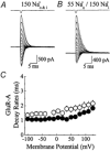
Figure 4
Figure 4. External ions do not affect AMPA receptor kinetics
A and B, Glu responses (10 mm Glu, 1 ms duration) evoked at a range of membrane potentials (−100 to + 125 mV, 15 mV increments) in external NaCl solutions of 150 mm (A) and 55 mm (B). Responses in each ionic condition were recorded from the same patch. Unlike kainate receptors, lowering NaCl concentration did not accelerate deactivation rates but produced a modest slowing of kinetics. The junction current (1 ms) recorded at the end of the experiment with an open patch electrode is shown above each trace. C, summary of the effect of external ions on GluR-A deactivation rates in 150 mm (n = 6, •) and 55 mm (n = 5, ○) external NaCl.
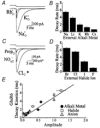
Figure 5
Figure 5. External cations and anions modulate amplitude and kinetics of kainate receptors
A, typical Glu responses (10 mm Glu, 50 ms duration, Vh=−20 mV) evoked in the same patch in 150 mm external Na, K or RbCl solutions. Replacement of external Na+ with K+ or Rb+ ions elicited a reversible, voltage-independent reduction in response amplitude and acceleration of desensitization kinetics. Upper trace shows the open tip current recorded at the end of the experiment. B, plot summarizing the effect of external alkali metal ions on the entry rate of GluR6 channels into desensitization. Statistical information is provided in Table 1. C, Glu responses (10 mm Glu, 50 ms duration, Vh=−20 mV) evoked in the same patch in the presence of 150 mm external chloride, nitrate or propionate sodium solutions. Similar to alkali metal ions, replacement of external chloride with nitrate or propionate elicited a reversible, voltage-independent reduction in response amplitude and acceleration of desensitization kinetics. Note that the agonist response observed in external Cl (marked by *) is significantly larger in amplitude and has been plotted at a different scale to permit comparison. Upper trace shows the open tip current. D, plot summarizing the effect of external halide ions on the entry rate of GluR6 channels into desensitization. Statistical information is provided in Table 2. E, plot comparing GluR6 response amplitude and desensitization kinetics under different external ion conditions. Data have been fit by linear regression analysis through the origin (continuous line, y = 4.94x, r = 0.83).
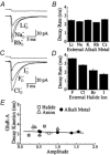
Figure 6
Figure 6. The effect of external cations and anions on AMPA receptor amplitude and kinetics
A, 10 mm Glu responses (50 ms duration, Vh=−20 mV) evoked in the same patch in the presence of 150 mm external Na, Rb or LiCl solutions. Replacement of external Na+ with Rb+ or Li+ ions elicited a reversible, voltage-independent increase or reduction in response amplitude respectively but did not affect desensitization kinetics. Upper trace shows the open tip current recorded at the end of the experiment. B, plot showing that external alkali metal ions do not influence the onset of GluR-A desensitization. Statistical information is provided in Table 1. C, typical Glu responses (10 mm Glu, 50 ms duration, Vh=−20 mV) evoked in the same patch in the presence of 150 mm external Cl, F or I Na solutions. Replacement of external Cl with F or I elicited a reversible, voltage-independent effect on desensitization kinetics and a reduction in response amplitude. Upper trace shows the open tip current. D, plot summarizing the effect of external halide ions on GluR-A desensitization rates. Statistical information is provided in Table 2. E, GluR-A response amplitude and desensitization kinetics in different external ion conditions are plotted for comparison. Data have been fit by linear regression analysis (continuous line, y =−0.22x + 2.6, r =−0.19).

Similar articles

Cited by

References

    1. Akk G, Auerbach A. Inorganic, monovalent cations compete with agonists for the transmitter binding site of nicotinic acetylcholine receptors. Biophysical Journal. 1996;70:2652–2658. - PMC - PubMed
    1. Antonov SM, Gmiro VE, Johnson JW. Binding sites for permeant ions in the channel of NMDA receptors and their effects on channel block. Nature Neuroscience. 1998;1:451–456. - PubMed
    1. Arai A, Silberg J, Kessler M, Lynch G. Effect of thiocyanate on AMPA receptor mediated responses in excised patches and hippocampal slices. Neuroscience. 1995;66:815–827. - PubMed
    1. Armstrong CM. Interaction of tetraethylammonium ion derivatives with the potassium channels of giant axons. Journal of General Physiology. 1971;58:413–437. - PMC - PubMed
    1. Armstrong N, Gouaux E. Mechanisms for activation and antagonism of an AMPA-sensitive glutamate receptor: crystal structures of the GluR2 ligand binding core. Neuron. 2000;28:165–181. - PubMed

Publication types

LinkOut - more resources