. 2002 Mar 15;539(Pt 3):903-11.
doi: 10.1113/jphysiol.2001.013282.

Excitability changes in human corticospinal projections to forearm muscles during voluntary movement of ipsilateral foot

Affiliations

Excitability changes in human corticospinal projections to forearm muscles during voluntary movement of ipsilateral foot

Fausto Baldissera et al. J Physiol. .

Abstract

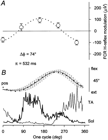
Excitability of the H-reflex in the relaxed flexor carpi radialis (FCR) muscle was tested during voluntary oscillations of the ipsilateral foot at five evenly spaced delays during a 600 ms cycle. In some experiments the H-reflex was conditioned by transcranial magnetic stimulation (TMS). With the hand prone, the amplitude of the FCR H-reflex was modulated sinusoidally with the same period as the foot oscillation, the modulation peak occurring in coincidence with contraction of the foot plantar-flexor soleus and the trough during contraction of the extensor tibialis anterior. When the H-reflex was facilitated by TMS at short latency (conditioning-test interval: -2 to -3.5 ms), the modulation was larger than that occurring with an unconditioned reflex of comparable size. This suggests that both the peripheral and the corticospinal components of the facilitated response were modulated in parallel. When the H-reflex was tested 40-60 ms after conditioning, i.e. during the cortical "silent period" induced by TMS, no direct effect was produced on the reflex size but the foot-associated modulation was deeply depressed. These results suggest that the reflex modulation may depend on activity fluctuations in the cortical motor area innervating the forearm motoneurones. It is proposed that when the foot is rhythmically oscillated, along with the full activation of the foot cortical area a simultaneous lesser co-activation of the forearm area produces a subliminal cyclic modulation of cervical motoneurones excitability. Should the two limbs be moved together, the time course of this modulation would favour isodirectional movements of the prone hand and foot, indeed the preferential coupling observed when hand and foot are voluntarily oscillated.


Figures

Figure 1
Figure 1. Schematic description of experimental procedure
Voluntary oscillations of the foot (uppermost trace) triggered a photocell at a fixed point of the movement cycle. After the third trigger, a PC-driven stimulator delivered an electric pulse to the median nerve, which evoked an H-reflex in the flexor xarpi radialis (FCR) muscle. The stimulus was timed, in random alternation, at one of five different delays (▴) dividing the cycle in equal fractions. In part of the trials, the H-reflex was conditioned by transcranial magnetic stimulation of the motor cortex. Rectified EMGs from tibialis anterior (TA) and soleus (Sol) muscles were also recorded (two lowermost traces). Box outlines the stimulation and recording period.
Figure 2
Figure 2. Cyclic modulation of FCR H-reflex during voluntary oscillation of ipsilateral foot
A, absolute deviations of the H-reflex size from its mean value (428 μV = 8 % Mmax; see Methods), occurring at five delays during voluntary oscillations of the foot. Each point represents the average (±s.e.m.) of 15 responses evoked at that delay. B illustrates the average record of the foot angular position (pos, uppermost continuous line, ±s.e.m.) and the rectified EMGs from Tibialis Anterior (TA, thick continuous trace) and Soleus (Sol, thin continuous trace) muscles. Dotted lines in A and B describe the sinusoidal functions that fitted the experimental data best (R2= 0.96 and 0.99, respectively). Period of the sine-wave function fitting the position record (π= 532 ms) was utilised both for fitting the reflex data and for normalising to 1 cycle the time scale of all parameters. The phase of the best-fit sinusoids for the movement and the H-reflex modulation was measured and their difference (ΔΦ) calculated. Positive values of ΔΦ indicate that the modulation sine wave advanced the movement sinusoid (plantar flexion, flex, upward).
Figure 3
Figure 3. Cyclic modulation of H-reflex, either unconditioned or conditioned by TMS, during voluntary foot oscillations
During foot oscillation, modulation of the unconditioned (○) and TMS-facilitated (•) H-reflexes follows a virtually identical course. The best-fit functions for the two sets of data (continuous and dotted lines) show a similar phase advance (ΔΦ) with respect to movement. Besides the period normalisation to one cycle, in each subject (6), data were normalised in size to the amplitude of the respective best-fit sine wave.
Figure 4
Figure 4. Size of CMAP evoked in FCR muscle by TMS stimulation of contralateral motor cortex is modulated during voluntary foot oscillations
A, the uppermost insets show the CMAPs evoked in one subject by TMS at 5 delays during the foot cycle (average of 15 responses ±s.e.m.). The lower graph shows modulation of the CMAP amplitude (•) and its best-fit sine wave (dotted line). Mean amplitude of CMAP = 423 μV. B, average records of the foot angular position (pos, upper continuous line, ±s.e.m.) and of the TA rectified EMG (lower continuous trace). Dotted line describes the best-fit function for movement (R2= 0.95) and ΔΦ is the phase difference between the best-fit sinusoids for the movement and the CMAP modulation. Cycle period (π) = 549 ms. Time calibration: major ticks = 25 ms, minor ticks = 5 ms.
Figure 5
Figure 5. Cyclic modulation of corticospinal facilitation of FCR H-reflex during voluntary foot oscillations
Amplitude of the H-reflex modulation at the delays corresponding to the peak (DEL1) and trough (DEL2) of the modulation cycle (symbolised by the continuous line) was larger when the reflex was facilitated by TMS (•) then when it was unconditioned (○). Conditioning-test interval ranged between −2 and −3.5 ms in the 5 subjects. Conditioned and unconditioned reflexes were tested in separate trials and their amplitude equalised between trials. The mean H-reflex size was around 5 % Mmax and not significantly different (paired t test) in the two conditions (174 μV for the unconditioned and 218 μV for the conditioned H reflex). Each couple of points, slightly shifted with respect to the others, refers to one subject. Mean amplitude of the modulation (▴ and ▵±s.e.m.) was significantly different in the two situations (P < 0.01, paired t test).
Figure 6
Figure 6. Cortical depression (silent period) suppresses cyclic H-reflex modulation associated with voluntary foot oscillations
H-reflex modulation at the peak (DEL1) and trough (DEL2) of the modulation cycle (○) was virtually suppressed when the reflex was evoked during the silent period induced by TMS delivered 40–60 ms in advance (•) with an intensity subliminal for evoking a CMAP. At these delays and intensities, TMS did not affect the H-reflex excitability; it was therefore unnecessary to correct the reflex size between trials. Mean values of the conditioned and unconditioned reflexes were not significantly different (paired t test) from each other (unconditioned = 231 μV; conditioned = 233 μV). Mean amplitude of the modulation was instead significantly different (P < 0.001, paired t test) in the two conditions (▴ and ▵±s.e.m.; s.e.m. bars are hidden by the symbols).

Similar articles

Cited by

References

    1. Aruin AS, Forrest WR, Latash ML. Anticipatory postural adjustments in conditions of postural instability. Electroencephalography & Clinical Neurophysiology. 1998;109:350–359. - PubMed
    1. Aruin AS, Latash ML. Directional specificity of postural muscles in feed-forward postural reactions during fast voluntary arm movements. Experimental Brain Research. 1995;103:323–332. - PubMed
    1. Aruin AS, Latash ML. Anticipatory postural adjustments during self-initiated perturbations of different magnitude triggered by a standard motor action. Electroencephalography & Clinical Neurophysiology. 1996;101:497–503. - PubMed
    1. Baldissera F, Borroni P, Cavallari P. Neural compensation for mechanical differences between hand and foot during coupled oscillations of the two segments. Experimental Brain Research. 2000;133:165–177. - PubMed
    1. Baldissera F, Borroni P, Cavallari P, Cerri G. Cyclic modulation of the excitability of resting forearm muscles is related to cyclic contraction of foot movers, not to movement. Pflügers Archiv. 2001;442:R7(4).

Publication types