. 2002 May;70(5):1305-17.
doi: 10.1086/340448. Epub 2002 Mar 15.

PKHD1, the polycystic kidney and hepatic disease 1 gene, encodes a novel large protein containing multiple immunoglobulin-like plexin-transcription-factor domains and parallel beta-helix 1 repeats

Affiliations

PKHD1, the polycystic kidney and hepatic disease 1 gene, encodes a novel large protein containing multiple immunoglobulin-like plexin-transcription-factor domains and parallel beta-helix 1 repeats

Luiz F Onuchic et al. Am J Hum Genet. 2002 May.

Abstract

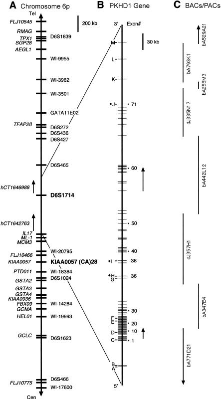
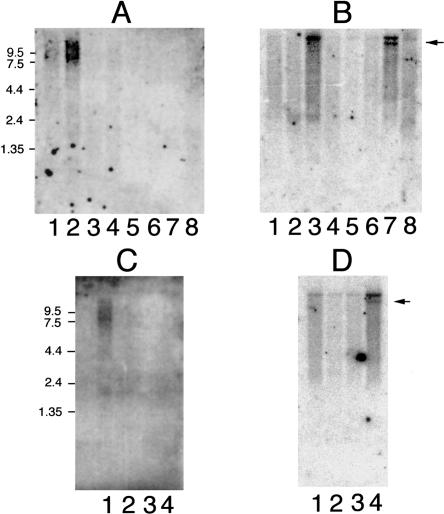
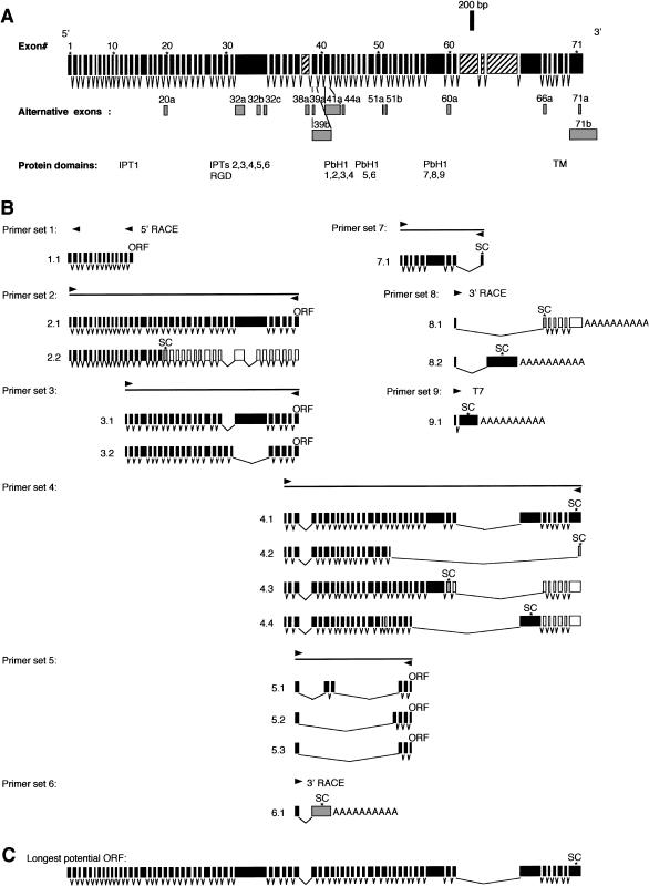
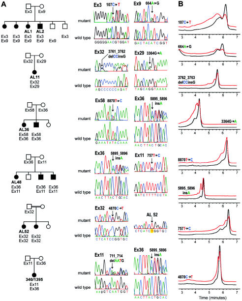
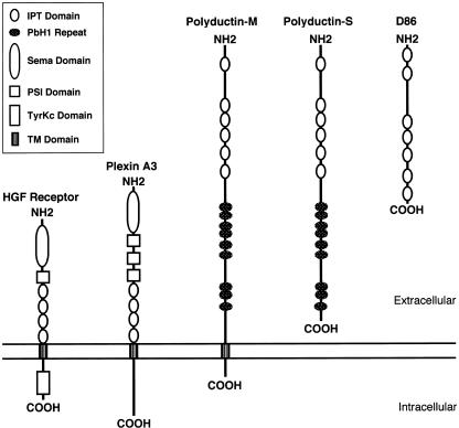
Autosomal recessive polycystic kidney disease (ARPKD) is a severe form of polycystic kidney disease that presents primarily in infancy and childhood and that is characterized by enlarged kidneys and congenital hepatic fibrosis. We have identified PKHD1, the gene mutated in ARPKD. PKHD1 extends over > or =469 kb, is primarily expressed in human fetal and adult kidney, and includes a minimum of 86 exons that are variably assembled into a number of alternatively spliced transcripts. The longest continuous open reading frame encodes a 4,074-amino-acid protein, polyductin, that is predicted to have a single transmembrane (TM)-spanning domain near its carboxyl terminus, immunoglobulin-like plexin-transcription-factor domains, and parallel beta-helix 1 repeats in its amino terminus. Several transcripts encode truncated products that lack the TM and that may be secreted if translated. The PKHD1-gene products are members of a novel class of proteins that share structural features with hepatocyte growth-factor receptor and plexins and that belong to a superfamily of proteins involved in regulation of cell proliferation and of cellular adhesion and repulsion.


Figures

Figure  A1
Figure A1
PKHD1 cDNA sequence encoding the longest potential reading frame. Colored bases establish the exon-exon boundaries: blue denotes the last base of the upstream exon; red, the first base of the downstream exon; green, the translation start site; pink, the stop codon.
Figure  A2
Figure A2
Amino acid sequence of polyductin encoded by the longest ORF of PKHD1. Blue denotes IPT domains; green, PbH1 repeats; red, TM domain; yellow, RGD domain.
Figure  1
Figure 1
Chromosomal localization and genomic organization of PKHD1. A, Schematic representation of chromosome 6p12. Currently known genes are identified on the far left (italics), and STSs/polymorphic markers are on the right. The closest flanking genetic markers that define the minimal PKHD1 interval are indicated (boldface). Not all of the 35 overlapping sets of expressed sequences described in the text are shown. B, Genomic organization of PKHD1. Exons identified by numbers have been shown to be part of PKHD1 transcripts. Letters indicate exons that belong to either hCT1642763 or hCT1646988 and that have not been confirmed by our analyses. The black diamond (♦) identifies, in hCT1642763 and hCT1646988, overlapping exons whose boundaries differ from those used in the present study. Arrows indicate the positions of the Gene Unit 6 and Gene Unit 442L12 transcripts described in the text. C, BACs and PACs sequenced by the Sanger Centre that cover the interval.
Figure  2
Figure 2
PKHD1 expression profile. A, Human adult multiple-tissue northern blot probed with PKHD1 exon 59: lane 1, pancreas; lane 2, kidney; lane 3, skeletal muscle; lane 4, liver; lane 5, lung; lane 6, placenta; lane 7, brain; lane 8, heart. B, Same blot as in panel A, probed with PKD1. The arrow indicates the position of a known splicing variant of PKD1. C, Human fetal multiple-tissue northern blot probed with PKHD1: lane, 1, kidney; lane 2, liver; lane 3, lung; lane 4, brain. D, Same blot as in panel C, probed with PKD1.
Figure  3
Figure 3
Structure of full-length PKHD1 and its splicing variants. A, Set of 71 nonoverlapping exons that spans the entire length of PKHD1 (upper row) and 15 additional overlapping exons that use different splice sites (gray boxes, lower row). Exons, which are not present in the cDNA that encodes the longest ORF, are indicated by hatched boxes. The position of important protein domains is indicated. B, Approximate location of each primer set used to amplify various cDNAs, with representative set of amplified products (below each schema). White boxes indicate noncoding exons in the corresponding transcripts while gray boxes identify exons with alternative boundaries (A). The templates used for each amplification are as follows: human adult kidney double-stranded cDNA for primer sets 1–4, 6, and 8; human kidney mRNA and total RNA for primer sets 5 and 7; human adult kidney cDNA library for primer set 9. “SC” indicates approximate location of stop codons, and “ORF” indicates that an open reading frame extends throughout the length of the fragment. C, Longest ORF identified by RT-PCR/cDNA amplification. This ORF is the composite sequence of products 2.1 and 4.1 of panel B and includes a total of 67 exons.
Figure  4
Figure 4
PKHD1 mutations in families studied. A, Representative family-segregation analyses of PKHD1 mutations for patients AL 1, AL 11, AL 36, AL 48, AL 52, and 340/1395 (see table 1). Sequence electropherograms showing wild-type and mutant sequences for amplicons containing the respective variants in each family are indicated on the right of each pedigree figure. Traces labeled “mutant” show heterozygous alterations in genomic PCR products. Segregation of the mutant allele (denoted by “Ex” followed by the exon number) is shown for each kindred studied. AL 1 has two missense changes, whereas AL 11, AL 36, and AL 48 each have a missense and a frameshifting mutation. Patient 340/1395 has two frameshifting mutations. AL 52 and her sibling, products of a consanguineous union, are homozygous for the Ex32 mutation (trace labeled AL 52). The Ex32 trace labeled “mutant” is from the heterozygous father. Black symbols denote affected individuals; white symbols denote unaffected individuals. B, Representative DHPLC profiles for several sequence variants in families with PKHD1 (see panel A and table 1). Black traces indicate control profiles; red traces indicate patient variant profiles (the red trace is displaced upward to facilitate comparison).
Figure  5
Figure 5
Structure of polyductin and related proteins. Multiple tandemly repeated IPT domains are common features of the group. Polyductin-M shares the general structure of the HGFR and plexin A3, in having a long extracellular domain, a single TM domain, and a short cytoplasmic carboxyl terminus, whereas polyductin-S is more like D86.

Comment in

References

Electronic-Database Information

    1. BLAST, http://www.ncbi.nlm.nih.gov/blast/
    1. Celera, http://public.celera.com/cds/login.cfm
    1. ExPASy Molecular Biology Server, http://ca.expasy.org/
    1. GenBank Overview, http://www.ncbi.nlm.nih.gov/Genbank/GenbankOverview.html (for sequences of all 86 exons and the composite cDNA with the longest ORF [accession number AF480064])
    1. GENSCAN, http://bioweb.pasteur.fr/seqanal/interfaces/genscan.html

References

    1. Burge C, Karlin S (1997) Prediction of complete gene structures in human genomic DNA. J Mol Biol 268:78–94 - PubMed
    1. Burset M, Guigo R (1996) Evaluation of gene structure prediction programs. Genomics 34:353–367 - PubMed
    1. Calvet JP (1993) Polycystic kidney disease: primary extracellular matrix abnormality or defective cellular differentiation? Kidney Int 43:101–108 - PubMed
    1. Cramer P, Larson CJ, Verdine GL, Muller CW (1997) Structure of the human NF kappaB p52 homodimer-DNA complex at 2.1 A resolution. EMBO J 16:7078–7090 - PMC - PubMed
    1. Eggermann T, Nothen MM, Propping P, Schwanitz G (1993) Molecular diagnosis of trisomy 18 using DNA recovered from paraffin embedded tissues and possible implications for genetic counselling Ann Genet 36:214–216 - PubMed

Publication types

MeSH terms

Associated data