

Leishmania major lipophosphoglycan modulates the phenotype and inhibits migration of murine Langerhans cells
- PMID: 11899433
- PMCID: PMC1783323
- DOI: 10.1046/j.1365-2567.2001.01333.x
Leishmania major lipophosphoglycan modulates the phenotype and inhibits migration of murine Langerhans cells
Abstract
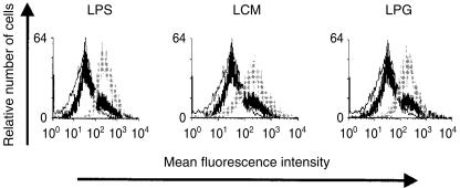
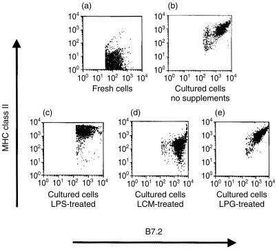
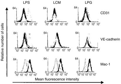
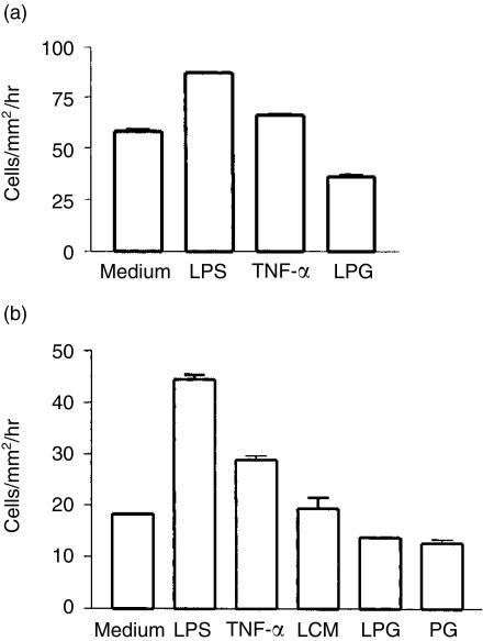
Langerhans cells (LC), members of the dendritic cell family, play a central role in the initiation and regulation of the immune response against the protozoan parasite Leishmania major. LC take up antigens in the skin and transport them to the regional lymph nodes for presentation to T cells. However, it is not known whether LC functions are modulated by parasite antigens. In the present study, we examined the effect of a major parasite surface molecule, L. major lipophosphoglycan (LPG), on the maturation of LC and their migratory properties. The results show that exposure to LPG did not affect the expression of major histocompatibility complex (MHC) class II and B7, but induced an up-regulation of CD25, CD31 and vascular endothelial (VE)-cadherin expression and a down-regulation of Mac-1 expression, by LC. Importantly, LPG treatment inhibited the migratory activity of LC, as it reduced their efflux from skin explants and their migration in transwell cultures. These results suggest that Leishmania LPG impairs LC migration out of the skin and thus may modulate their immunostimulatory functions, which require LC translocation from skin to lymph nodes.
Figures
Similar articles
-
[Leishmania major lipophosphoglycan modulates the expression of receptors involved in parasite internalization in skin Langerhans cells].Acta Cient Venez. 2002;53(3):218-24. Acta Cient Venez. 2002. PMID: 12658871 Spanish.
-
Leishmania lipophosphoglycan reduces monocyte transendothelial migration: modulation of cell adhesion molecules, intercellular junctional proteins, and chemoattractants.J Immunol. 1998 Feb 15;160(4):1857-65. J Immunol. 1998. PMID: 9469447
-
Systemic treatment with anti-CD40 antibody stimulates Langerhans cell migration from the skin.Clin Exp Immunol. 2002 Sep;129(3):519-26. doi: 10.1046/j.1365-2249.2002.01909.x. Clin Exp Immunol. 2002. PMID: 12197894 Free PMC article.
-
Langerhans cells transport Leishmania major from the infected skin to the draining lymph node for presentation to antigen-specific T cells.Eur J Immunol. 1993 Jul;23(7):1595-601. doi: 10.1002/eji.1830230730. Eur J Immunol. 1993. PMID: 8325337
-
Accessory and adhesion molecules expressed on murine epidermal Langerhans cells and the modulation by cytokines.J Dermatol Sci. 1998 May;20(1):14-20. doi: 10.1016/s0923-1811(99)00006-7. J Dermatol Sci. 1998. PMID: 10342744 Review.
Cited by
-
Dectin-1 Positive Dendritic Cells Expand after Infection with Leishmania major Parasites and Represent Promising Targets for Vaccine Development.Front Immunol. 2018 Feb 26;9:263. doi: 10.3389/fimmu.2018.00263. eCollection 2018. Front Immunol. 2018. PMID: 29535708 Free PMC article.
-
Modulation of dendritic cell responses by parasites: a common strategy to survive.J Biomed Biotechnol. 2010;2010:357106. doi: 10.1155/2010/357106. Epub 2010 Feb 24. J Biomed Biotechnol. 2010. PMID: 20204070 Free PMC article. Review.
-
Targeting Leishmania major Antigens to Dendritic Cells In Vivo Induces Protective Immunity.PLoS One. 2013 Jun 26;8(6):e67453. doi: 10.1371/journal.pone.0067453. Print 2013. PLoS One. 2013. PMID: 23840706 Free PMC article.
-
Innate immune activation and subversion of Mammalian functions by leishmania lipophosphoglycan.J Parasitol Res. 2012;2012:165126. doi: 10.1155/2012/165126. Epub 2012 Feb 22. J Parasitol Res. 2012. PMID: 22523640 Free PMC article.
-
Role of the C-type lectins DC-SIGN and L-SIGN in Leishmania interaction with host phagocytes.Immunobiology. 2005;210(2-4):185-93. doi: 10.1016/j.imbio.2005.05.013. Immunobiology. 2005. PMID: 16164025 Free PMC article. Review.
References
-
- Liew FY, O'Donnell CA. Immunology of leishmaniasis. Adv Parasitol. 1993;32:161–259. - PubMed
-
- Alexander J, Russell DG. The interaction of Leishmania species with macrophages. Adv Parasitol. 1992;31:175–254. - PubMed
-
- Moll H. Epidermal Langerhans cells are critical for immunoregulation of cutaneous leishmaniasis. Immunol Today. 1993;14:383–7. - PubMed
-
- Fruth U, Solioz N, Louis JA. Leishmania major interferes with antigen presentation by infected macrophages. J Immunol. 1993;150:1857–64. - PubMed
-
- Kaye PM, Rogers NJ, Curry AJ, Scott JC. Deficient expression of co-stimulatory molecules on Leishmania-infected macrophages. Eur J Immunol. 1994;24:2850–4. - PubMed
Publication types
MeSH terms
Substances
LinkOut - more resources
Full Text Sources
Research Materials
